
Research Article 
 Creative Commons, CC-BY
Peptide-Based Therapeutics in Human Male Fertility: Molecular Mechanisms, Translational Evidence, and Clinical Prospects
*Corresponding author:Jonathan RT Lakey, PhD, MSM, Professor Emeritus Departments of Surgery and Biomedical Engineering University of California Irvine, USA.
Received:May 01, 2026; Published:May 12, 2026
DOI: 10.34297/AJBSR.2026.31.004012
Abstract
Male infertility accounts for approximately 40–50% of infertility cases globally and represents a growing public health challenge associated with declining sperm quality, endocrine dysfunction, and environmental stressors. Conventional treatments, including hormonal therapy, antioxidant supplementation, and assisted reproductive technologies, often address symptoms rather than the underlying molecular pathology responsible for impaired spermatogenesis. Peptide-based therapeutics have recently emerged as promising biologic regulators capable of restoring reproductive homeostasis at the molecular level. Bioactive peptides, short amino-acid sequences acting as regulatory signalling molecules, modulate critical pathways governing endocrine regulation, mitochondrial bioenergetics, oxidative stress control, angiogenesis, and tissue regeneration. Evidence from mammalian translational models demonstrates that peptide therapies enhance spermatogenic activity, improve sperm motility and mitochondrial function, restore steroidogenic signalling, and promote gonadal tissue repair. Particularly promising are mitochondrial-targeted peptides and organ-specific peptide complexes that regulate steroidogenesis via pathways involving StAR, PGC-1α, NRF1, and TFAM. Emerging nano-delivery technologies further enhance peptide stability, tissue targeting, and bioavailability. Integration of peptide therapeutics with precision medicine approaches may enable individualized fertility restoration strategies. This review synthesizes current mechanistic, experimental, and translational evidence supporting peptide therapeutics as a next-generation modality in human male reproductive medicine.
Keywords:male infertility, peptide therapeutics, mitochondrial peptides, spermatogenesis, oxidative stress, HPG axis, reproductive biotechnology
Introduction
Male reproductive health represents a critical determinant of human fertility, population sustainability, and long-term demographic stability [1,2]. Infertility affects approximately 10–15% of couples globally, and male factors are implicated in nearly half of these cases, either as a sole cause or in combination with female reproductive disorders [3]. Recent epidemiologic analyses suggest that the burden of male infertility is increasing worldwide, reflecting both improved diagnostic recognition and a growing prevalence of underlying biological risk factors. In parallel, large-scale meta- analyses have documented a substantial decline in sperm concentration and total sperm counts over the past several decades. Levine and colleagues reported that sperm counts among men from Western countries decreased by more than 50% between 1973 and 2011, a trend that has continued into the present decade [2]. These observations have generated considerable concern within the reproductive health community, raising questions regarding the environmental, metabolic, and molecular determinants of declining male fertility.
Male infertility encompasses a heterogeneous spectrum of clinical conditions that impair the production, maturation, or function of spermatozoa. These disorders are conventionally categorized according to abnormalities in semen parameters, including oligospermia (reduced sperm concentration), azoospermia (complete absence of sperm), asthenozoospermia (reduced sperm motility), teratozoospermia (abnormal sperm morphology), and endocrine-related infertility associated with hormonal dysregulation [4]. Although these phenotypic categories are widely used in clinical andrological practice, they represent downstream manifestations of complex underlying biological processes [5]. Increasing evidence indicates that male infertility frequently results from the interplay of endocrine disturbances, oxidative stress, mitochondrial dysfunction, inflammatory signalling, genetic abnormalities, and environmental exposures [6]. Understanding how these mechanisms converge to disrupt spermatogenesis and sperm function remains an area of active investigation.
At the physiological level, male reproductive function is governed by the tightly regulated Hypothalamic–Pituitary–Gonadal (HPG) axis, which coordinates endocrine signaling required for spermatogenesis and androgen production [6]. Pulsatile secretion of Gonadotropin-Releasing Hormone (GnRH) from the hypothalamus stimulates the pituitary to release Luteinizing Hormone (LH) and Follicle-Stimulating Hormone (FSH). LH acts primarily on Leydig cells to stimulate testosterone synthesis, whereas FSH regulates Sertoli cell function and supports germ-cell maturation within the seminiferous tubules [7]. Disruption of this regulatory network can impair testosterone production, compromise Sertoli cell support of developing germ cells, and ultimately lead to defective spermatogenesis [7]. Endocrine abnormalities such as hypogonadotropic hypogonadism, altered feedback regulation of gonadotropins, or disturbances in androgen signaling therefore represent important contributors to male reproductive dysfunction [8]. Beyond endocrine regulation, emerging evidence highlights the central role of cellular metabolism and mitochondrial function in determining sperm quality and fertilization capacity. Mitochondria located in the sperm midpiece generate Adenosine Triphosphate (ATP) required for flagellar movement and progressive motility [9]. Impairment of mitochondrial oxidative phosphorylation can therefore directly compromise sperm motility and fertilization competence [10]. Moreover, mitochondria serve as a major source of Reactive Oxygen Species (ROS), which at physiological levels participate in essential processes such as sperm capacitation and acrosome reaction but become deleterious when produced in excess [9]. Elevated oxidative stress within the male reproductive tract can induce lipid peroxidation of sperm membranes, DNA fragmentation, and apoptosis of germ cells [11]. Indeed, oxidative stress is now recognized as one of the most prevalent molecular contributors to male infertility, implicated in up to 80% of idiopathic infertility cases [12]. Environmental and lifestyle factors further amplify these biological vulnerabilities. Exposure to endocrine-disrupting chemicals, heavy metals, air pollutants, and agricultural pesticides has been associated with impaired semen quality and reduced reproductive potential [13]. Similarly, metabolic disorders such as obesity, diabetes mellitus, and metabolic syndrome have been linked to altered hormonal signalling, increased oxidative stress, and disruption of testicular function [14]. These multifactorial influences suggest that male infertility should be understood not as a single disease entity but rather as a complex systems-level disorder arising from the intersection of endocrine, metabolic, environmental, and genetic determinants. This systems-level perspective reframes male infertility as a failure of coordinated biological networks rather than isolated dysfunction within a single pathway. Disruptions in endocrine signaling, mitochondrial bioenergetics, redox homeostasis, and immune regulation do not occur independently but instead interact dynamically to destabilize the testicular microenvironment required for spermatogenesis. Consequently, therapeutic strategies that target only downstream manifestations may be insufficient to restore physiological reproductive function. In this context, bioactive peptides have emerged as candidate modulators of these interconnected systems, functioning not as conventional supplements but as signaling molecules capable of interfacing with endogenous regulatory pathways. By influencing receptor-mediated signaling, transcriptional networks, and cellular stress responses, peptide-based interventions offer a mechanistically integrated approach to restoring reproductive homeostasis at the molecular level (Table 1).

Table 1:Causes of Male Infertility.
Despite advances in diagnostic evaluation, therapeutic options for male infertility remain limited. Current management strategies primarily focus on correcting endocrine abnormalities or circumventing impaired sperm function through Assisted Reproductive Technologies (ART), including In Vitro Fertilization (IVF) and intracytoplasmic sperm injection [15]. Although these approaches have improved reproductive outcomes for many couples, they often bypass rather than correct the underlying biological processes responsible for impaired fertility [16]. Moreover, ART procedures are associated with substantial financial cost, psychological burden, and potential health risks, highlighting the need for alternative therapeutic strategies that directly target the molecular drivers of male reproductive dysfunction [17].
In recent years, advances in molecular biology, regenerative medicine, and peptide biotechnology have stimulated growing interest in bioactive peptides as modulators of reproductive physiology. Peptides are short amino-acid chains that function as signaling molecules capable of regulating cellular metabolism, gene expression, immune responses, and endocrine pathways [18]. Unlike traditional pharmacologic agents that broadly affect systemic physiology, peptides often exhibit high receptor specificity and act through physiologic signaling networks, enabling more targeted modulation of biological processes [19]. In the context of reproductive biology, peptide-mediated signaling has been implicated in diverse processes including steroidogenesis, mitochondrial regulation, angiogenesis, and cellular stress responses [20].
Particularly intriguing are mitochondrial-derived peptides and organ-specific regulatory peptides, which have been shown to influence cellular energy metabolism, oxidative balance, and tissue regeneration. Experimental studies suggest that such peptides can enhance mitochondrial biogenesis, reduce oxidative damage, and modulate transcriptional pathways involved in cellular resilience [21,22]. These mechanisms are highly relevant to male reproductive function, as spermatogenesis and sperm motility are energetically demanding processes that depend on efficient mitochondrial metabolism and tightly regulated redox homeostasis. Moreover, peptide signaling pathways may influence endocrine regulation within the HPG axis and support the maintenance of the testicular microenvironment necessary for germ-cell development [23].
The integration of peptide therapeutics into reproductive medicine therefore represents a promising avenue for addressing the molecular drivers of male infertility. By targeting pathways involved in cellular energy metabolism, oxidative stress regulation, and endocrine signaling, peptides have the potential to restore physiological homeostasis within the male reproductive system rather than merely compensating for downstream functional deficits. Although clinical data remain limited, accumulating evidence from translational and experimental studies supports the concept that peptide-based interventions may enhance spermatogenic activity, improve sperm quality, and promote reproductive tissue repair. Given the growing prevalence of male infertility and the limitations of current therapeutic approaches, there is a pressing need to better understand the molecular mechanisms underlying reproductive dysfunction and to explore innovative treatment strategies capable of restoring reproductive health.
Pathophysiology of Male Infertility
Disruption of GnRH pulsatility alters the temporal dynamics of pituitary gonadotropin secretion, leading to impaired Leydig cell steroidogenesis and compromised Sertoli cell support of germ-cell development. upstream regulatory pathways play a critical role in maintaining GnRH neuronal activity. Kisspeptin-expressing neurons within the hypothalamus act as central modulators of GnRH release, integrating metabolic and hormonal signals to coordinate reproductive function [24]. Perturbations in kisspeptin signaling have been directly associated with hypogonadotropic states and reduced gonadotropin output. In parallel, systemic metabolic cues, e.g., leptin and insulin, modulate hypothalamic function, linking energy homeostasis to reproductive capacity. Dysregulation of these pathways, particularly in obesity and metabolic syndrome, can suppress GnRH secretion and downstream androgen production [25].
Hypothalamic–Pituitary–Gonadal Axis Dysfunction
At the peripheral level, circulating Sex Hormone–Binding Globulin (SHBG) critically determines the fraction of bioavailable testosterone accessible to target tissues [24]. Age-related increases in SHBG contribute to a progressive decline in free testosterone despite relatively stable total testosterone concentrations. Longitudinal studies indicate that total testosterone levels decline by approximately 1–2% annually in aging males, with a more pronounced reduction in bioavailable androgen due to rising SHBG concentrations [26]. This endocrine shift has significant implications for spermatogenesis, as intratesticular testosterone concentrations are essential for maintaining germ-cell maturation. Collectively, these findings highlight that HPG axis dysfunction extends beyond hormonal deficiency to encompass disruptions in neuroendocrine regulation, metabolic signaling, and hormone bioavailability [27]. Such disturbances may arise from congenital hypogonadotropic hypogonadism, metabolic disorders, systemic illness, or environmental endocrine disruptors that interfere with steroidogenic signaling pathways [8]. In addition to classical reproductive hormones, emerging biomarkers such as Anti-Müllerian Hormone (AMH) and insulin-like peptide 3 (INSL3) provide important insights into Sertoli and Leydig cell function, respectively [28]. Altered concentrations of these biomarkers reflect disturbances within the testicular microenvironment and have been associated with impaired spermatogenic capacity and reduced semen quality [29] (Table 2).
Alterations in AMH, INSL3, and testosterone levels are therefore increasingly recognized as clinically relevant biomarkers of testicular dysfunction and endocrine dysregulation in infertile men [30,31].
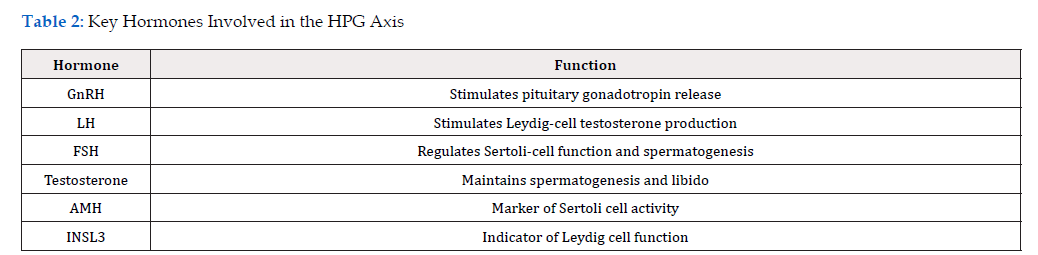
Table 2:Key Hormones Involved in the HPG Axis
Oxidative Stress
Excessive Reactive Oxygen Species (ROS) production disrupts redox homeostasis and induces lipid peroxidation of sperm membranes, compromising membrane fluidity and functional integrity [32]. his process is accompanied by the accumulation of oxidative biomarkers, including 8-hydroxy-2′-deoxyguanosine (8-OHdG), which reflects oxidative DNA damage, and Malondialdehyde (MDA), a byproduct of lipid peroxidation. Concurrent depletion of intracellular antioxidant systems, particularly reduced glutathione, further exacerbates cellular vulnerability to oxidative injury [11,33]. These molecular alterations impair sperm motility, disrupt chromatin integrity, and reduce fertilization potential [11]. The sperm plasma membrane contains exceptionally high concentrations of polyunsaturated fatty acids that are highly susceptible to lipid peroxidation [34]. In addition, mature sperm cells possess minimal cytoplasmic antioxidant defenses due to the extensive cytoplasmic reduction that occurs during spermiogenesis [35]. Importantly, oxidative stress does not act in isolation but is closely coupled to mitochondrial dysfunction. Excess ROS generated during oxidative phosphorylation can damage mitochondrial membranes and mitochondrial DNA, initiating a self-perpetuating cycle of oxidative injury. This interplay between ROS generation and mitochondrial impairment represents a central mechanism linking environmental stressors, metabolic dysfunction, and declining sperm quality [33].
Mitochondrial Dysfunction
Sperm function is critically dependent on mitochondrial oxidative phosphorylation, which is mediated by electron transport chain complexes I–V embedded within the inner mitochondrial membrane [36]. Emerging evidence indicates that mitochondrial dysfunction in male infertility is characterized by a bidirectional feedback loop in which ROS-induced mitochondrial damage further enhances ROS generation, amplifying cellular injury. This cycle contributes to progressive declines in sperm motility and viability. In addition, mitochondrial dysfunction has been linked to broader hallmarks of cellular aging, including telomere shortening and replicative senescence, which may further impair germ-cell renewal capacity [37].
Inflammation and Immune Dysregulation
Chronic inflammatory signaling within the male reproductive tract disrupts the specialized microenvironment required for spermatogenesis. Activation of nuclear factor kappa B (NF-κB) pathways in response to infection, metabolic stress, or tissue injury leads to increased production of pro-inflammatory cytokines, including TNF-α, IL-1β, and IL-6. These cytokines not only induce germ-cell apoptosis but also interfere with Sertoli cell function and disrupt intercellular communication within the seminiferous epithelium [38,39]. Inflammatory signaling also exerts direct effects on mitochondrial function, as cytokine-mediated pathways can impair electron transport chain activity and increase mitochondrial ROS production. This interaction further reinforces the link between inflammation and oxidative stress in male infertility.
In addition, disruption of the blood–testis barrier compromises immune privilege within the testes, exposing germ-cell antigens to the systemic immune system and promoting the formation of antisperm antibodies. Such immune-mediated responses can impair sperm function and reduce fertilization capacity, highlighting the importance of immune homeostasis in maintaining reproductive health. These interconnected mechanisms illustrate the multifactorial nature of male infertility and highlight the importance of therapeutic strategies capable of restoring physiological homeostasis across multiple biological systems. Understanding these pathogenic pathways provides a mechanistic foundation for exploring novel molecular interventions aimed at improving male reproductive health.
Table 3 highlights molecular pathways implicated in the pathophysiology of male infertility, integrating endocrine, metabolic, oxidative, mitochondrial, and inflammatory mechanisms. Disruptions within the HPG axis alter gonadotropin signaling and androgen bioavailability, while oxidative stress and mitochondrial dysfunction converge to impair sperm bioenergetics and genomic integrity. Inflammatory signaling further exacerbates these effects through cytokine-mediated damage and breakdown of immune privilege within the testes. The interconnected nature of these pathways highlights the systems-level basis of male infertility and underscores the need for therapeutic strategies capable of targeting multiple biological domains simultaneously. These integrated mechanisms provide a conceptual framework for the targeted application of peptide-based therapeutics discussed in subsequent sections (Table 3).
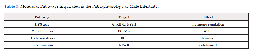
Table 3:Molecular Pathways Implicated in the Pathophysiology of Male Infertility.
Biological Basis of Peptide Therapeutics
Peptides function as endogenous signaling molecules that regulate cellular physiology through highly specific molecular interactions. Their biological activity is mediated through diverse signaling modalities, including activation of G-Protein–Coupled Receptors (GPCRs), such as melanocortin and neuropeptide receptors, engagement of receptor tyrosine kinases, and direct intracellular interactions with transcriptional regulators [40]. Through these mechanisms, peptides influence key cellular processes including metabolic regulation, stress-response signaling, and gene expression networks that govern cellular adaptation and survival [41]. Unlike conventional pharmacologic agents that often act through broad systemic effects, peptides typically operate within physiological signaling cascades, allowing targeted modulation of discrete molecular pathways. In reproductive biology, such signaling is particularly relevant given the need for precise coordination between endocrine, metabolic, and cellular processes. Peptide-mediated regulation of intracellular signaling pathways enables modulation of transcription factors, mitochondrial function, and inflammatory responses that collectively shape the testicular microenvironment [42]. However, the therapeutic application of peptides is constrained by inherent pharmacokinetic limitations. Native peptides are highly susceptible to proteolytic degradation, resulting in short biological half-lives and reduced systemic stability. Rapid enzymatic breakdown within plasma and tissues limits their bioavailability and necessitates the development of specialized delivery systems to preserve biological activity [43]. Advances in peptide stabilization, including chemical modification, encapsulation within nanocarriers, and sustained-release formulations, have therefore become critical for enabling effective clinical translation. These strategies not only enhance peptide stability but also facilitate targeted delivery to specific tissues, improving therapeutic precision while minimizing off-target effects [44]. Because peptides are derived from naturally occurring amino-acid sequences, they generally demonstrate high receptor specificity and reduced off-target effects [44]. Their structural similarity to endogenous signaling molecules also contributes to favourable safety profiles, including relatively low systemic toxicity and minimal immunogenicity when compared with larger protein therapeutics [45]. Furthermore, peptides can often be designed to mimic physiological signaling pathways rather than disrupt them, allowing targeted modulation of biological processes that are dysregulated in disease states [46]. Spermatogenesis is a highly energy-dependent and tightly regulated developmental process that requires coordinated endocrine signaling, efficient mitochondrial metabolism, adequate vascular supply, and protection from oxidative and inflammatory stress [47].
Endocrine Modulation
Through influencing neuroendocrine signaling by modulating the release of GnRH from hypothalamic neurons or altering the sensitivity of pituitary gonadotroph cells to GnRH stimulation, peptide- mediated signaling can enhance the secretion of LH and follicle- stimulating hormone, the two principal pituitary hormones responsible for regulating testicular function [23]. By influencing these hormonal pathways, peptides may help restore endocrine balance in individuals with functional disruptions of the HPG axis, [48] particularly relevant in conditions characterized by impaired gonadotropin secretion, age-related hormonal decline, or metabolic disorders that interfere with androgen signaling. Importantly, peptide-mediated endocrine modulation differs from conventional hormone replacement strategies because it acts upstream within physiological signaling pathways, potentially allowing restoration of endogenous hormone production rather than reliance on exogenous hormone supplementation [49].
Mitochondrial Bioenergetic Restoration
Mitochondria are central to sperm physiology because they generate the ATP required for flagellar movement and progressive motility [50]. Dysfunctional mitochondria reduce ATP availability and can impair sperm motility, viability, and fertilization potential [36,51]. Certain bioactive peptides, including mitochondrial-derived peptides, have been shown to influence mitochondrial metabolism by stabilizing mitochondrial membrane potential, improving electron transport chain efficiency, and reducing the accumulation of ROS generated during oxidative phosphorylation [41]. These effects help maintain cellular redox balance and prevent oxidative damage to mitochondrial DNA and membrane structures [11]. Peptides have also been shown to activate transcriptional pathways involved in mitochondrial biogenesis and metabolic adaptation [52]. Key regulatory proteins such as peroxisome proliferator–activated receptor gamma coactivator 1-alpha (PGC-1α), Nuclear Respiratory Factor 1 (NRF1), and mitochondrial Transcription Factor A (TFAM) coordinate the replication of mitochondrial DNA and the synthesis of mitochondrial proteins required for oxidative phosphorylation [53]. Activation of these pathways can enhance mitochondrial density and metabolic capacity within cells. In germ cells and spermatozoa, improved mitochondrial bioenergetics may translate into enhanced sperm motility, improved resistance to oxidative stress, and greater overall reproductive competence [50].
Angiogenesis and Microvascular Support
Adequate vascularization is essential for maintaining the metabolic and endocrine functions of reproductive tissues [54]. The testes possess an extensive microvascular network that supplies oxygen, nutrients, and hormonal signals required for germ-cell development and steroidogenesis. Impairment of testicular blood flow has been associated with reduced spermatogenic activity and compromised testosterone production [55]. Peptide signaling pathways can influence angiogenesis by modulating the activity of VEGF and other angiogenic mediators that regulate endothelial- cell proliferation and capillary formation [56]. Enhanced angiogenic signaling promotes the formation of new blood vessels and improves tissue perfusion within reproductive organs. Improved microvascular circulation may facilitate more efficient nutrient delivery, waste removal, and endocrine communication within the testicular microenvironment [57]. In this way, peptides that stimulate angiogenic pathways may indirectly support spermatogenesis by optimizing the metabolic and hormonal conditions required for germ-cell development.
Paracrine Regeneration and Secretome Signaling
An additional mechanism through which peptides influence reproductive physiology involves paracrine signaling derived from stem-cell secretomes. Mesenchymal Stem Cells (MSCs) and other regenerative cell populations release a complex mixture of cytokines, growth factors, extracellular vesicles, and bioactive peptides that collectively regulate tissue repair and immune responses [58]. Increasing evidence suggests that many of the regenerative effects attributed to stem-cell therapies arise not from direct cell engraftment but from the signaling molecules present within this secretome [59]. Peptide components of the secretome can modulate inflammatory signaling, promote tissue regeneration, and inhibit fibrotic remodeling within damaged tissues [59]. Within the male reproductive system, these paracrine signals may help restore the specialized cellular microenvironment required for spermatogenesis by promoting Sertoli-cell survival, enhancing vascular stability, and suppressing inflammatory responses that disrupt germ-cell development [60]. Because secretome-derived peptides can replicate many of the regenerative effects of MSC without the complexities associated with cellular transplantation, they represent an attractive therapeutic strategy for restoring tissue homeostasis in reproductive disorders. Taken together, these mechanisms illustrate the diverse ways in which peptide signaling may influence male reproductive physiology. By modulating endocrine communication, mitochondrial metabolism, vascular integrity, and regenerative signaling pathways, peptide-based interventions offer a biologically integrated approach to restoring the cellular environment necessary for normal spermatogenesis and sperm function.
Mitochondrial Peptides
Mitochondrial-Derived Peptides (MDP) represent an emerging class of bioactive molecules that originate from short open reading frames encoded within mitochondrial DNA. MDP function as signaling mediators linking mitochondrial metabolic status to nuclear gene expression and cellular stress responses [61]. In recent years, MDP such as humanin, MOTS-c (Mitochondrial Open Reading Frame of the 12S rRNA-c), and small humanin-like peptides have been shown to regulate metabolic homeostasis, oxidative resilience, and cellular survival pathways [62]. Unlike classical mitochondrial proteins involved directly in oxidative phosphorylation, MDP act as endocrine-like signaling molecules capable of influencing cellular function at both local and systemic levels [63]. Within reproductive biology, mitochondrial peptides are particularly relevant because germ-cell maturation and sperm motility are highly dependent on mitochondrial energy metabolism [63]. Experimental studies demonstrate that MDP can enhance mitochondrial respiration efficiency, maintain mitochondrial membrane potential, and regulate redox homeostasis under conditions of metabolic stress [21]. These peptides also influence intracellular signaling pathways associated with mitochondrial biogenesis and metabolic adaptation, including activation of AMP-Activated Protein Kinase (AMPK) and downstream transcriptional regulators that coordinate mitochondrial replication and repair [64]. Through these mechanisms, MDPs help maintain cellular ATP availability while limiting the accumulation of reactive oxygen species generated during oxidative phosphorylation. Such effects suggest that mitochondrial peptide signaling may contribute to maintaining germ-cell integrity and supporting the energetically demanding process of spermatogenesis. Because mitochondrial dysfunction is strongly implicated in male infertility and reproductive aging, therapies targeting mitochondrial signaling pathways represent a promising strategy for restoring cellular homeostasis within the testicular microenvironment.
Organ-Specific Peptides/b>
Organ-derived peptide fractions have been investigated for their ability to modulate tissue-specific cellular processes. Organ- specific peptide signaling has been studied in multiple physiological contexts, including immune regulation, endocrine signaling, and tissue regeneration [65]. In reproductive medicine, peptides derived from endocrine or reproductive tissues may exert regulatory effects on gene expression programs that govern cellular differentiation, steroidogenesis, and tissue repair [66]. Experimental evidence indicates that certain peptide complexes can modulate transcriptional activity in a tissue-specific manner, influencing the expression of genes involved in cellular metabolism, stress responses, and structural integrity [65]. This phenomenon is thought to occur through interactions between peptide ligands and intracellular transcriptional regulators that control gene expression within target cells. Within the male reproductive system, organ-derived peptides may support the specialized cellular interactions that occur between Sertoli cells, Leydig cells, and developing germ cells [67]. Multi-factorial peptide complexes have been developed to combine peptides derived from multiple endocrine and reproductive organs in order to address the hierarchical nature of HPG axis regulation [26]. These peptide complexes are proposed to modulate gene expression through interactions with nuclear and cytoplasmic signaling pathways, influencing transcriptional programs involved in steroidogenesis, cellular metabolism, and tissue repair. Experimental and observational studies suggest that such peptides may exert epigenetic-like regulatory effects, promoting restoration of cellular function without directly replacing hormones or cells. This gene-regulatory mechanism provides a biologically plausible basis for their reported ability to support endocrine balance and tissue homeostasis within reproductive systems.
Nano-Peptide Delivery Systems
Although peptides possess significant therapeutic potential, their clinical utility is often limited by rapid degradation, limited tissue penetration, and short systemic half-life. Advances in nanotechnology have therefore led to the development of peptide delivery systems designed to enhance stability, bioavailability, and target specificity. Nanoparticle-based delivery platforms allow peptides to be encapsulated within protective carriers that shield them from enzymatic degradation while facilitating controlled release at target tissues [68]. Nano-formulated peptides can be engineered to interact selectively with specific cell types or molecular targets through surface modifications that enhance receptor binding or cellular uptake. These systems may improve intracellular peptide delivery by enabling peptides to cross cellular membranes more efficiently than conventional formulations [68]. Additionally, nanocarriers can prolong peptide circulation time within the bloodstream, thereby increasing the probability of reaching target tissues [68]. Nano-peptide technologies may enable targeted delivery of bioactive peptides to reproductive organs or to specific cell populations within the testes. Improved delivery efficiency could enhance the therapeutic effects of peptides while minimizing systemic exposure and potential off-target effects. As nanotechnology continues to evolve, the integration of peptide therapeutics with advanced delivery systems may substantially expand the clinical applicability of peptide-based regenerative strategies (Table 4).

Table 4:Mechanisms of Peptide Therapy.
Translational Evidence
The translational development of peptide-based therapeutics in male reproductive medicine is supported by a growing body of experimental, ex vivo, and early clinical evidence, although the field remains at a relatively early stage of clinical validation. Collectively, these studies suggest that peptide-mediated modulation of mitochondrial function, oxidative stress, and endocrine signaling may exert measurable effects on sperm quality and reproductive physiology. However, the strength of evidence varies substantially across experimental systems, necessitating careful distinction between mechanistic plausibility and clinically demonstrated efficacy [69].
At the preclinical level, multiple studies have demonstrated that bioactive peptides, particularly those derived from mitochondrial signaling pathways, influence cellular metabolism and stress resilience. Mitochondrial-derived peptides such as MOTS-c and humanin have been shown to regulate AMP-Activated Protein Kinase (AMPK) signaling, enhance insulin sensitivity, and attenuate oxidative stress through modulation of ROS production and mitochondrial membrane stability. These effects are highly relevant to spermatogenesis, a process characterized by substantial demands and sensitivity to redox imbalance. Experimental models indicate that improved mitochondrial efficiency is associated with enhanced ATP availability, reduced apoptotic signaling, and preservation of germ-cell integrity, thereby providing a mechanistic basis for the application of peptide therapeutics in male fertility.
Ex vivo and cell-based studies further support the role of peptide- rich biological systems in maintaining sperm function under stress conditions. Conditioned media derived from MSCs, which contains a complex secretome of cytokines, growth factors, extracellular vesicles, and peptides, has been shown to improve sperm motility, viability, and mitochondrial respiration following cryopreservation [70]. These findings are particularly notable in the context of assisted reproductive technologies, where oxidative stress and mitochondrial dysfunction contribute to reduced sperm quality. The beneficial effects observed in these systems are thought to be mediated by restoration of mitochondrial membrane potential, reduction of lipid peroxidation, and activation of intracellular survival pathways [62]. Importantly, such studies highlight that the therapeutic activity may reside not in cellular transplantation per se, but in the peptide-rich signaling milieu of the secretome, thereby supporting the development of cell-free peptide-based interventions.
Emerging clinical and observational data provide preliminary support for the translational relevance of these mechanisms, although robust randomized evidence remains limited. Investigations conducted within regenerative medicine frameworks, including those reported by the European Wellness Biomedical Group, have explored the use of organ-specific peptide complexes and mitochondrial-derived peptide formulations targeting components of the HPG axis [71]. In a longitudinal cohort evaluating biohormonal revitalization therapy in men with hypogonadal features, administration of multi-organ peptide formulations derived from hypothalamic, pituitary, adrenal, and testicular tissues was associated with significant increases in serum testosterone levels, rising from baseline values in the hypogonadal range (~280-300ng/dL) to mid-physiological levels (~490-506 ng/dL) after four months of treatment, with partial maintenance of these gains at one-year follow-up [72]. These hormonal changes were accompanied by reported improvements in libido, energy levels, and overall well-being, suggesting potential endocrine and systemic benefits of peptide- based interventions. However, these findings are derived from a non-randomized cohort with limited sample size and absence of a control group, restricting causal inference. Complementary reports from regenerative and integrative medicine studies further suggest that peptide-based therapies, particularly when combined with stem-cell–derived products or exosome-rich formulations, may enhance vascular, metabolic, and endocrine parameters relevant to male reproductive health. For example, integrative treatment frameworks incorporating peptide signaling, hormonal optimization, and regenerative modalities have demonstrated improvements in sexual function and systemic health markers in men with erectile dysfunction, a condition closely linked to endothelial dysfunction, metabolic disease, and impaired reproductive capacity. Erectile dysfunction, increasingly recognized as a manifestation of systemic endothelial and metabolic dysfunction, shares key pathophysiological pathways with male infertility. These findings provide preliminary translational support for the hypothesis that peptide- based interventions may modulate reproductive physiology.
Despite these promising observations, several critical limitations must be acknowledged. Much of the available clinical evidence is derived from small, non-randomized cohorts or observational studies lacking appropriate control groups, thereby limiting causal inference. In addition, heterogeneity in peptide formulations, dosing regimens, and delivery methods complicates cross-study comparisons and prevents standardization of therapeutic protocols. Importantly, few studies have evaluated hard reproductive endpoints such as pregnancy rates, live birth outcomes, or long-term offspring health, which remain essential for clinical translation in fertility medicine.
Taken together, the current body of evidence supports a biologically plausible and mechanistically grounded role for peptide therapeutics in modulating key pathways underlying male infertility, particularly those related to mitochondrial bioenergetics, oxidative stress, and endocrine regulation. However, the field now requires rigorously designed randomized controlled trials, standardized peptide characterization, and long-term outcome studies to determine whether the cellular and physiological benefits observed in preclinical and early clinical studies can be translated into meaningful improvements in human fertility.
Safety and Regulatory Considerations
While peptide-based therapeutics are often regarded as biologically compatible due to their derivation from endogenous amino- acid sequences, their safety profile in the context of reproductive medicine requires careful and systematic evaluation. The assumption of inherent safety based on physiological origin may not fully account for the pharmacologic effects observed when peptides are administered at supraphysiologic concentrations, delivered through non-native routes, or combined into multi-component formulations. Consequently, a rigorous assessment of potential risks is essential for clinical translation. A consideration is the potential for immunogenicity, particularly with repeated administration or structurally modified peptides designed to enhance stability. Although short peptides are generally less immunogenic than larger protein biologics, immune responses can still occur, especially when peptides are delivered via nanoparticle carriers or conjugated to stabilizing molecules. Such responses may alter therapeutic efficacy or, in rare cases, induce inflammatory or hypersensitivity reactions [73].
In addition, off-target receptor activation represents a significant concern given the pleiotropic nature of peptide signaling pathways. Many peptide receptors, including G-protein–coupled receptors, are expressed across multiple tissues, raising the possibility that exogenous peptides may activate unintended signaling cascades outside the reproductive system. This is particularly relevant for peptides involved in metabolic or neuroendocrine regulation, where systemic effects could influence cardiovascular, neurological, or metabolic function. Another critical safety consideration is the risk of endocrine overstimulation. Peptide-mediated modulation of the HPG axis may enhance endogenous hormone production; however, excessive or prolonged stimulation could disrupt normal feedback mechanisms. Dysregulated gonadotropin or androgen levels may lead to altered spermatogenic dynamics, hormonal imbalances, or downstream effects on other endocrine axes. Careful titration of dosing regimens and monitoring of hormonal biomarkers are therefore essential to minimize the risk of iatrogenic endocrine disruption.
Furthermore, the reproductive safety profile of peptide therapeutics remains incompletely characterized, particularly with respect to long-term outcomes. While short-term improvements in semen parameters or hormonal levels are frequently reported, there is limited evidence regarding the impact of peptide interventions on fertilization success, embryo development, and offspring health. Given that germ cells carry genetic and epigenetic information transmitted to subsequent generations, any therapeutic intervention affecting spermatogenesis warrants thorough evaluation for potential heritable effects. From a regulatory perspective, peptide therapeutics occupy a complex position between small-molecule drugs and biologics, requiring tailored frameworks for approval. Standard requirements include pharmacokinetic and pharmacodynamic characterization, toxicology studies, and evaluation of tissue distribution. However, in reproductive medicine, additional endpoints must be incorporated into clinical trial design. The heterogeneity of peptide formulations, particularly multi-organ or secretome-derived products, further complicates standardization and quality control. Collectively, these considerations underscore the need for rigorously designed clinical studies, standardized manufacturing processes, and long-term safety monitoring. While peptide therapeutics offer substantial promise as targeted modulators of reproductive physiology, their integration into clinical practice must be guided by comprehensive evaluation of both efficacy and safety to ensure responsible and effective application in male fertility treatment (Figure 1).

Figure 1:Schematic representation of the multi-level mechanisms by which peptide-based therapeutics modulate male reproductive function. The central panel illustrates the anatomical and functional integration of the Hypothalamic-Pituitary-Gonadal (HPG) axis with the testicular microenvironment, highlighting the endocrine and cellular pathways governing spermatogenesis. Surrounding panels depict four principal domains of peptide action. Endocrine modulation (upper left) demonstrates peptide-mediated regulation of hypothalamic GnRH release and downstream pituitary secretion of Luteinizing Hormone (LH) and Follicle- Stimulating Hormone (FSH), resulting in enhanced testosterone production and support of spermatogenesis. Mitochondrial restoration (upper right) illustrates the effects of mitochondrial-derived peptides on cellular bioenergetics, including increased ATP production, reduced Reactive Oxygen Species (ROS) accumulation, and activation of mitochondrial biogenesis pathways. Vascular support (lower left) highlights angiogenic peptide signaling that promotes Vascular Endothelial Growth Factor (VEGF) activation, increased blood flow, and improved nutrient delivery to reproductive tissues. Paracrine regeneration (lower right) depicts the role of peptide-rich stem-cell secretomes in mediating anti-inflammatory effects, immune modulation, and tissue repair within the testicular microenvironment. The lower panel summarizes key translational outcomes associated with these mechanisms, including improved sperm motility, enhanced mitochondrial function, increased gamete viability, and restoration of endocrine balance. Collectively, the figure illustrates the systems-level integration of peptide signaling pathways in restoring male reproductive homeostasis.
Conclusion
Despite promising advances, several critical questions remain regarding the clinical role of peptide therapeutics in male infertility. Rigorous randomized controlled trials will be required to determine whether peptide-based therapies can significantly improve fertility outcomes in human populations. In addition, further investigation is needed to characterize peptide pharmacodynamics, optimize dosing strategies, and identify biomarkers that predict therapeutic response. Future research should also explore potential synergistic interactions between peptide therapeutics and other regenerative strategies, including stem-cell secretome therapies, mitochondrial-targeted antioxidants, and metabolic modulators. Such combination approaches may prove particularly valuable in treating complex cases of idiopathic infertility where multiple pathological mechanisms are present. Male infertility is a multifactorial condition arising from complex interactions between endocrine regulation, cellular metabolism, oxidative stress, and immune signaling. Peptide-based therapeutics represent a promising new class of biological modulators capable of influencing many of these pathways simultaneously. By targeting mitochondrial metabolism, endocrine signaling networks, and regenerative pathways, peptides offer a biologically integrated approach to restoring reproductive homeostasis.
Although clinical translation remains in early stages, advances in peptide biotechnology, nanomedicine, and precision diagnostics are rapidly expanding the therapeutic potential of peptide signaling. Continued interdisciplinary research will be essential to determine how these innovations can be safely and effectively integrated into clinical strategies for the treatment of male infertility
Competing Interests
None.
Acknowledgement
None.
References
- De Jonge CJ, Barratt CLR, Aitken RJ, et al. (2024) Current global status of male reproductive health. Hum Reprod Open 2024(2):
- Levine H, Jørgensen N, Martino-Andrade A, et al. (2017) Temporal trends in sperm count: a systematic review and meta-regression analysis. Hum Reprod Update 23(6): 646-659.
- Agarwal A, Baskaran S, Parekh N, et al. (2021) Male infertility. Lancet 397(10271): 319-333.
- Krausz C, Riera-Escamilla A (2018) Genetics of male infertility. Nat Rev Urol 15(6): 369-384.
- Wyrwoll MJ, van der Heijden GW, Krausz C, et al. (2024) Improved phenotypic classification of male infertility to promote discovery of genetic causes. Nat Rev Urol 21(2): 91-101.
- Skakkebaek NE, Rajpert-De Meyts E, Buck Louis GM, et al. (2016) Male Reproductive Disorders and Fertility Trends: Influences of Environment and Genetic Susceptibility. Physiol Rev 96(1): 55-97.
- Walker WH (2011) Testosterone signaling and the regulation of spermatogenesis. Spermatogenesis 1(2): 116-120.
- Sengupta P, Dutta S, Karkada IR, Chinni SV (2021) Endocrinopathies and Male Infertility. Life 12(1):
- Irigoyen P, Pintos-Polasky P, Rosa-Villagran L, Skowronek MF, Cassina A, et al. (2022) Mitochondrial metabolism determines the functional status of human sperm and correlates with semen parameters. Front Cell Dev Biol 10:
- Amaral A, Lourenço B, Marques M, Ramalho-Santos J (2013) Mitochondria functionality and sperm quality. Reproduction 146(5): R163-174.
- Wang Y, Fu X, Li H (2025) Mechanisms of oxidative stress-induced sperm dysfunction. Front Endocrinol 16:
- Pavuluri H, Bakhtiary Z, Panner Selvam MK, Hellstrom WJG (2024) Oxidative Stress-Associated Male Infertility: Current Diagnostic and Therapeutic Approaches. Medicina (Mex) 60(6):
- Stavros S, Kathopoulis N, Moustakli E, et al. (2025) Endocrine-Disrupting Chemicals and Male Infertility: Mechanisms, Risks, and Regulatory Challenges. J Xenobiotics 15(5):
- Zhang W, Tong L, Jin B, Sun D (2025) Diabetic testicular dysfunction and spermatogenesis impairment: mechanisms and therapeutic prospects. Front Endocrinol 16:
- Abdul Rahman H, Mazlan B, Amdani SN (2025) A Scoping Review of Male Fertility Assessment and Treatment Across Historical and Contemporary Paradigms. Andrology 15:
- Emokpae MA, Brown SI (2021) Effects of lifestyle factors on fertility: practical recommendations for modification. Reprod Fertil 2(1): R13-R26.
- Gül M, Russo GI, Kandil H, et al. (2024) Male Infertility: New Developments, Current Challenges, and Future Directions. World J Mens Health 42(3): 502-517.
- Han Y, Zhang Y, Li H, Ma Z, Wang Y (2025) Peptide Drug: Design and Clinical Applications. MedComm 6(8):
- Rossino G, Marchese E, Galli G, et al. (2023) Peptides as Therapeutic Agents: Challenges and Opportunities in the Green Transition Era. Molecules 28(20):
- Lu Z, Chai Q, Dai W, et al. (2025) Mitochondrial homeostasis restoring peptide-drug conjugates with ROS-responsive NO releasing ability for targeted therapy of myocardial infarction. J Nanobiotechnology 23(1):
- Ran Y, Guo Z, Zhang L, et al. (2025) Mitochondria‑derived peptides: Promising microproteins in cardiovascular diseases (Review). Mol Med Rep 31(5):
- Miller B, Kim SJ, Kumagai H, et al. (2020) Peptides derived from small mitochondrial open reading frames: Genomic, biological, and therapeutic implications. Exp Cell Res 393(2):
- Li L, Lin W, Wang Z, et al. (2024) Hormone Regulation in Testicular Development and Function. Int J Mol Sci 25(11):
- Lei T, Yang Y, Yang WX. (2025) Luteinizing Hormone Regulates Testosterone Production, Leydig Cell Proliferation, Differentiation, and Circadian Rhythm During Spermatogenesis. Int J Mol Sci 26(8):
- Zhou R, Wu J, Liu B, et al. (2019) The roles and mechanisms of Leydig cells and myoid cells in regulating spermatogenesis. Cell Mol Life Sci CMLS 76(14): 2681-2695.
- Klokol D, Chan MKS, Nillenthiran L, et al. (2019) Biohormonal revitalization therapy from the perspective of biological regenerative medicine: the evaluation of premature menopause and andropause treatment outcomes in longitudinal cohort study 10(4).
- Tsutsumi R, Webster NJG (2009) GnRH pulsatility, the pituitary response and reproductive dysfunction. Endocr J 56(6): 729-737.
- Lukač NB, Škrlep M, Poklukar K, et al. (2026) INSL3 Expression in Leydig Cells is a Biomarker for Immunocastration in Boars: Transcriptional Evidence. Andrology 14(3): 782-795.
- Zikopoulos A, Christopoulos P, Kalampokas T, et al. (2026) Oxidative Stress and Inflammatory Biomarkers in Male Infertility: A Narrative Review of Diagnostic Value and Clinical Integration. Diagnostics 16(4): 527.
- Chong YH, Pankhurst MW, McLennan IS (2017) The Testicular Hormones AMH, InhB, INSL3, and Testosterone Can Be Independently Deficient in Older Men. J Gerontol A Biol Sci Med Sci 72(4): 548-553.
- Sansone A, Kliesch S, Isidori AM, Schlatt S (2019) AMH and INSL3 in testicular and extragonadal pathophysiology: what do we know? Andrology 7(2): 131-138.
- Sengupta P, Pinggera GM, Calogero AE, Agarwal A (2024) Oxidative stress affects sperm health and fertility-Time to apply facts learned at the bench to help the patient: Lessons for busy clinicians. Reprod Med Biol 23(1): e12598.
- Walke G, Gaurkar SS, Prasad R, Lohakare T, Wanjari M (2023) The Impact of Oxidative Stress on Male Reproductive Function: Exploring the Role of Antioxidant Supplementation. Cureus 15(7):
- Evans EPP, Scholten JTM, Mzyk A, et al. (2021) Male subfertility and oxidative stress. Redox Bio 46: 102071.
- Kowalczyk A (2022) The Role of the Natural Antioxidant Mechanism in Sperm Cells. Reprod Sci 29(5): 1387-1394.
- Mai Z, Yang D, Wang D, et al. (2024) A narrative review of mitochondrial dysfunction and male infertility. Transl Androl Urol 13(9): 2134-2145.
- Yemeliyanova M, Chan MKS, Wong MBF, Klokol, Dmytro (2024) Unexplained infertility: a fresh look at the old problem and the novel therapeutic options of its treatment. Obstet Gynecol Int J 15(1).
- Perez CV, Theas MS, Jacobo PV, Jarazo Dietrich S, Guazzone VA, et al. (2013) Dual role of immune cells in the testis: Protective or pathogenic for germ cells? Spermatogenesis 3(1): e23870.
- Yao PL, Lin YC, Richburg JH (2009) TNF alpha-mediated disruption of spermatogenesis in response to Sertoli cell injury in rodents is partially regulated by MMP2. Biol Reprod 80(3): 581-589.
- Wang L, Wang N, Zhang W, Xurui Cheng, Zhibin Yan et al. (2022) Therapeutic peptides: current applications and future directions. Signal Transduct Target Ther 7(1): 48.
- Akbarian M, Khani A, Eghbalpour S, Uversky VN (2022) Bioactive Peptides: Synthesis, Sources, Applications, and Proposed Mechanisms of Action. Int J Mol Sci 23(3): 1445.
- Bliss SP, Navratil AM, Xie J, Roberson MS (2010) GnRH signaling, the gonadotrope and endocrine control of fertility. Front Neuroendocrinol 31(3): 322-340.
- Cho YY, Kim S, Kim P, Min Jeong Jo, SongE Park, et al. (2025) G-Protein-Coupled Receptor (GPCR) Signaling and Pharmacology in Metabolism: Physiology, Mechanisms, and Therapeutic Potential. Biomolecules 15(2): 291.
- Li Q, Chao W, Qiu L (2025) Therapeutic peptides: chemical strategies fortify peptides for enhanced disease treatment efficacy. Amino Acids 57(1): 25.
- Liu M, Svirskis D, Proft T, Jacelyn Loh, Naibo Yin, et al. (2025) Progress in peptide and protein therapeutics: Challenges and strategies. Acta Pharm Sin B 15(12): 6342-6381.
- Zhang Z, Svensson KJ (2025) Discovery of peptides as key regulators of metabolic and cardiovascular crosstalk. Cell Rep 24; 44(6): 115836.
- Kaltsas A, Kyrgiafini MA, Mamuris Z, Chrisofos M, Sofikitis N (2026) Spermatogenesis Beyond DNA: Integrated RNA Control of the Epitranscriptome and Three-Dimensional Genome Architecture. Curr Issues Mol Biol 48(1):
- AcevedoRodriguez A, Kauffman AS, Cherrington BD, Borges CS, Roepke TA, et al. (2018) Emerging insights into hypothalamic-pituitary-gonadal axis regulation and interaction with stress signalling. J Neuroendocrinol 30(10): e12590.
- Kolodziejski PA, Pruszynska Oszmalek E, Wojciechowicz T, Maciej Sassek, Natalia Leciejewska, et al. (2021) The Role of Peptide Hormones Discovered in the 21st Century in the Regulation of Adipose Tissue Functions. Genes 12(5): 756.
- Xu Z, Yan Q, Zhang K, Ying Lei, Chen Zhou, et al. (2025) Mitochondrial Regulation of Spermatozoa Function: Metabolism, Oxidative Stress and Therapeutic Insights. Animal (Basel) 15(15): 2246.
- Vahedi Raad M, Firouzabadi AM, Tofighi Niaki M, Henkel R, Fesahat F (2024) The impact of mitochondrial impairments on sperm function and male fertility: a systematic review. Reprod Biol Endocrinol 22(1):83.
- Wan W, Zhang L, Lin Y, Xiuqing Rao, Xifeng Wang, et al. (2023) Mitochondria-derived peptide MOTS-c: effects and mechanisms related to stress, metabolism and aging. J Transl Med 21(1): 36.
- Cao L, Li Y, Smirnov A, Ramouna Voshtani, Tingting Wang, et al. (2025) PGC-1α: key regulator of mitochondrial biogenesis and cellular differentiation in metabolic and regenerative tissues. Cell Biosci 16(1): 9.
- Stucker S, De Angelis J, Kusumbe AP (2021) Heterogeneity and Dynamics of Vasculature in the Endocrine System During Aging and Disease. Front Physiol 9:12: 624928.
- Reyes JG, Farias JG, Henriquez Olavarrieta S, Eva Madrid, Mario Parraga, et al. (2012) The hypoxic testicle: physiology and pathophysiology. Oxid Med Cell Longev 2012: 2012: 929285.
- Kim M, Park S, Kim S, Seo J, Roh S (2025) A Novel Cell-Penetrating Peptide-Vascular Endothelial Growth Factor Small Interfering Ribonucleic Acid Complex That Mediates the Inhibition of Angiogenesis by Human Umbilical Vein Endothelial Cells and in an Ex Vivo Mouse Aorta Ring Model. Biomater Res 7: 29: 0120.
- Yi J, Lee S, Ryu C, Kim G, Kim J, et al. (2025) Enhanced Angiogenic Potential of Electrically Stimulated Human Adipose-Derived Mesenchymal Stem Cells (MSCs) for Ischemic Tissue Regeneration. Med Comm (2020) 6(9): e70352.
- Gonzalez Gonzalez A, Garcia Sanchez D, Dotta M, Rodriguez Rey JC, Perez Campo FM (2020) Mesenchymal stem cells secretome: The cornerstone of cell-free regenerative medicine. World J Stem Cells 12(12): 1529-1552.
- Trigo CM, Rodrigues JS, Camoes SP, Sola S, Miranda JP (2025) Mesenchymal stem cell secretome for regenerative medicine: Where do we stand? J Adv Res 70: 103-124.
- Ruthig VA, Lamb DJ (2022) Updates in Sertoli Cell-Mediated Signaling During Spermatogenesis and Advances in Restoring Sertoli Cell Function. Front Endocrinol 13: 897196.
- Walker BR, Moraes CT (2022) Nuclear-Mitochondrial Interactions. Biomolecules 12(3): 427.
- Wan W, Zhang L, Lin Y, Xiuqing Rao, Xifeng Wang, et al. (2023) Mitochondria-derived peptide MOTS-c: effects and mechanisms related to stress, metabolism and aging. J Transl Med 21(1): 36.
- Mohtashami Z, Singh MK, Salimiaghdam N, Ozgul M, Kenney MC (2022) MOTS-c, the Most Recent Mitochondrial Derived Peptide in Human Aging and Age-Related Diseases. Int J Mol Sci 23(19): 11991.
- Li C, Reif MM, Craige SM, Kant S, Keaney JF (2016) Endothelial AMPK activation induces mitochondrial biogenesis and stress adaptation via eNOS-dependent mTORC1 signaling. Nitric Oxide Biol Chem 1: 55-56: 45-53.
- Slivka JP, Bauer C, Younsi A, Wong MBF, Chan MKS, et al. (2024) Exploring the Molecular Tapestry: Organ-Specific Peptide and Protein Ultrafiltrates and Their Role in Therapeutics. Int J Mol Sci 25(5): 2863.
- Janssens Y, Wynendaele E, Vanden Berghe W, Bart De Spiegeleer (2019) Spiegeleer B Peptides as epigenetic modulators: therapeutic implications. Clin Epigenetics 11(1): 101.
- Hofmann MC, McBeath E (2022) Sertoli Cell-Germ Cell Interactions Within the Niche: Paracrine and Juxtacrine Molecular Communications. Front Endocrinol 13: 897062.
- Zhang X, Li X, Zhao Y, Zheng Q, Wu Q, et al. (2022) Nanocarrier system: An emerging strategy for bioactive peptide delivery. Front Nutr 9: 1050647.
- Bosler JS, Davies KP, Neal Perry GS (2014) Peptides in seminal fluid and their role in infertility: a potential role for opiorphin inhibition of neutral endopeptidase activity as a clinically relevant modulator of sperm motility: a review. Reprod Sci 21(11): 1334-1340.
- Nafchi HG, Azizi Y, Halvaei I (2024) Effect of Conditioned Medium from Human Adipose-Derived Mesenchymal Stem Cells on Human Sperm Quality During Cryopreservation. Reprod Sci 31(6): 1586-1592.
- European Wellness Official Website. Peptide Therapy Publications and Case Studies. https://european-wellness.eu/category/publications/peptides/
- Klokol D, Wong MBF, Chernykh V, et al. (2026) Modern Management of Erectile Dysfunction: An Integrative and Regenerative Therapeutic Framework. SSP Mod Pharm Med 6(1).
- Lee C, Zeng J, Drew BG, Tamer Sallam, Alejandro Martin Montalvo, et al. (2015) The mitochondrial-derived peptide MOTS-c promotes metabolic homeostasis and reduces obesity and insulin resistance. Cell Metab 21(3): 443-454.




 We use cookies to ensure you get the best experience on our website.