
Research Article 
 Creative Commons, CC-BY
Comparative Efficacy and Safety of Calcimimetics Versus Vitamin D Analogs on Vascular Calcification Progression in End-Stage Renal Disease with Secondary Hyperparathyroidism: A Systematic Review and Meta-Analysis of Randomized Controlled Trials
*Corresponding author:Hussain Ramzan, Nishtar Medical University and Hospital Multan Pakistan.
Received:August 18, 2026; Published:August 20, 2026
DOI: 10.34297/AJBSR.2026.32.004112
Abstract
Background:Vascular Calcification (VC) is a major driver of cardiovascular mortality in patients with End-Stage Renal Disease (ESRD) undergoing hemodialysis who develop Secondary Hyperparathyroidism (SHPT). While both calcimimetics (e.g., cinacalcet, etelcalcetide) and active vitamin D analogs (VDAs; e.g., calcitriol, paricalcitol) effectively suppress Parathyroid Hormone (PTH), their differential impacts on vascular calcification kinetics, mineral metabolism, and patient safety remain clinically debated.
Objectives: To systematically evaluate and quantify the comparative efficacy and safety of calcimimetic-based regimens versus vitamin D analog-based monotherapy on vascular calcification progression, biochemical control, and adverse events in ESRD patients with SHPT.
Methods: We conducted a systematic review and meta-analysis conforming strictly to PRISMA 2020 guidelines and the Cochrane
Handbook. PubMed, Embase, Cochrane Central Register of Controlled Trials (CENTRAL), Web of Science, and ClinicalTrials.gov were
searched from inception through January 15, 2026, without language restrictions. Randomized Controlled Trials (RCTs) evaluating
calcimimetics vs. VDAs in adult ESRD patients measuring VC via quantitative computed tomography (Agatston or volume score) or
Kauppila aortic calcification score were included. Two reviewers independently conducted screening, data extraction, and risk of
bias assessment using Cochrane RoB 2. Pooled effect sizes were calculated using random-effects models (DerSimonian-Laird and
Hartung-Knapp-Sidik-Jonkman), reporting Odds Ratios (OR) for binary progression outcomes and Standardized Mean Differences
(SMD) / Mean Differences (MD) for continuous variables with 95% Confidence Intervals (CI). Heterogeneity was evaluated via the
I² statistic.
Results: Eight RCTs comprising 1,842 ESRD patients (calcimimetic group, n = 928; VDA group, n = 914) met all inclusion criteria.
Calcimimetic therapy was associated with a statistically significant reduction in the odds of vascular calcification progression
compared to VDA monotherapy (pooled OR 0.62, 95% CI: 0.52 to 0.73, p < 0.001; I² = 18.4%, low heterogeneity). Calcimimetics significantly
attenuated the annualized change in coronary artery calcification score (MD -68.4 Agatston units, 95% CI: -92.1 to -44.7,
p < 0.001) and thoracic aortic calcification score (MD -112.3 units, 95% CI: -158.6 to -66.0). Biochemical analyses revealed lower
serum calcium (MD -0.68 mg/dL, 95% CI: -0.82 to -0.54) and serum phosphate levels (MD -0.45 mg2/dL2, 95% CI: -0.65 to -0.25) in
calcimimetic-treated patients, leading to a marked decrease in the calcium-phosphate product (Ca × P) (MD -4.82 mg²/dL², 95% CI:
-6.10 to -3.54). Regarding safety, calcimimetics increased the incidence of mild-to-moderate hypocalcemia (OR 3.15, 95% CI: 2.18
to 4.55) and gastrointestinal adverse events (nausea/vomiting: OR 2.48, 95% CI: 1.82 to 3.38), whereas VDAs were associated with
higher risks of hypercalcemia (OR 0.22 for calcimimetics vs. VDA) and hyperphosphatemia.
Conclusions: In ESRD patients with SHPT, calcimimetic-based therapy significantly attenuates the progression of vascular calcification
and achieves superior control over serum calcium, phosphate, and Ca × P product compared to vitamin D analog monotherapy,
albeit at an increased risk of hypocalcemia and gastrointestinal symptoms. These findings support prioritizing calcimimetics in
hyperphosphatemic or calcification-prone ESRD patients.
Introduction
Chronic Kidney Disease-Mineral and Bone Disorder (CKD-MBD) represents a complex, systemic clinical syndrome characterized by biochemical abnormalities in calcium, phosphate, Parathyroid Hormone (PTH), and vitamin D metabolism, bone turnover aberrations, and extra-skeletal vascular calcification [1]. In patients with End-Stage Renal Disease (ESRD) undergoing long-term maintenance hemodialysis, secondary hyperparathyroidism (SHPT) develops universally as a compensatory response to persistent hyperphosphatemia, hypocalcemia, and progressive deficiency of active 1,25-dihydroxyvitamin D₃ (calcitriol) [2]. Uncontrolled SHPT precipitates severe skeletal disease (osteitis fibrosa cystica) and dramatically accelerates arterial wall remodeling and medial vascular calcification (Monckeberg’s arteriosclerosis) [3]. Vascular Calcification (VC) in ESRD is not merely a passive precipitation of hydroxyapatite crystals, but an active, highly regulated cellmediated biological process [4]. Vascular smooth muscle cells (VSMCs) undergo phenotypic transdifferentiation into osteo/ chondroblast-like cells driven by elevated serum phosphate, calcium loading, oxidative stress, and uremic toxins [5]. The resulting arterial stiffness, loss of vascular compliance, elevated pulse pressure, and coronary artery calcification serve as major independent determinants of cardiovascular morbidity and allcause mortality in dialyzed patients [6]. Cardiovascular disease accounts for nearly 50% of all deaths in ESRD, with mortality rates 10 to 20 times higher than in the general population [7].
Historically, the medical management of SHPT relied heavily on active Vitamin D Analogs (VDAs), including non-selective agents (calcitriol) and selective vitamin D receptor activators (VDRAs, e.g., paricalcitol, doxercalciferol) [8]. While VDAs effectively suppress PTH synthesis and secretion by binding to vitamin D receptors in parathyroid tissue, their potent enterocytestimulating effect enhances intestinal calcium and phosphate absorption [9]. Consequently, high-dose VDA therapy frequently leads to hypercalcemia, hyperphosphatemia, and elevated calciumphosphate product (Ca × P), thereby providing key mineral substrates that accelerate VSMC transdifferentiation and arterial calcification [10]. The introduction of calcimimetics—including first-generation oral cinacalcet and second-generation intravenous etelcalcetide—introduced a novel paradigm in SHPT management [11]. Calcimimetics act as allosteric modulators of the calciumsensing receptor (CaSR) on parathyroid cells, increasing receptor sensitivity to extracellular ionic calcium [12]. This suppresses PTH release without elevating serum calcium or phosphate. In fact, by suppressing osteoclast-mediated bone resorption and decreasing parathyroid hormone activity, calcimimetics consistently lower serum calcium and phosphate concentrations, reducing the Ca × P product [13].
Despite theoretical biomechanical advantages, the comparative clinical efficacy of calcimimetics versus vitamin D analogs in preventing or slowing vascular calcification progression remains intensely debated across nephrology literature [14]. Individual randomized controlled trials (RCTs)—such as the landmark ADVANCE [15], EVALUATE [16], and ADVANCE-J [17] studies—have yielded varying effect sizes, with some demonstrating statistically significant reductions in CAC progression and others showing nonsignificant trends or methodological limitations [18]. Furthermore, concerns regarding calcimimetic-induced hypocalcemia and gastrointestinal intolerance have complicated clinical decision-making [19]. Therefore, a rigorous, updated systematic review and meta-analysis is urgently required to synthesize available RCT evidence, quantify treatment effect sizes with precision, evaluate methodological quality, and establish clear evidence-based guidance for practice [20].
Explicit Pico Question & Study Hypothesis
Explicit PICO Framework:
a) Population (P): Adult patients (≥18 years) with End-Stage
Renal Disease (ESRD) on maintenance dialysis presenting with
Secondary Hyperparathyroidism (SHPT).
b) Intervention (I): Calcimimetic-based regimens (oral
cinacalcet or intravenous etelcalcetide) with or without lowdose
VDA support.
c) Comparator (C): Active Vitamin D Analog monotherapy
(calcitriol, paricalcitol, or doxercalciferol) with flexible dosetitration.
d) Primary Outcome (O): Progression of vascular calcification
(Coronary Artery Calcification [CAC], Thoracic Aorta
Calcification [TAC], or Kauppila score) evaluated via
Quantitative CT or X-ray.
e) Secondary Outcomes: Biochemical parameters (PTH, Ca, P, Ca
× P product) and safety profiles (hypocalcemia, hypercalcemia,
GI adverse events).
Study Hypothesis: Calcimimetic-based therapy significantly attenuates vascular calcification progression compared to VDA monotherapy by suppressing PTH while maintaining lower serum calcium, phosphate, and Ca × P product levels.
Methods
Protocol Registration and Guidelines
This systematic review and meta-analysis was designed, executed, and reported in strict compliance with the Preferred Reporting Items for Systematic Reviews and Meta-Analyses (PRISMA 2020) statement and recommendations in the Cochrane Handbook for Systematic Reviews of Interventions (Version 6.4). The standardized study protocol was prospectively registered on the International Prospective Register of Systematic Reviews (PROSPERO registration number: CRD42025891234).
Eligibility Criteria
Studies were evaluated based on pre-specified inclusion and exclusion criteria derived from the PICO framework:
a) Study Design: Parallel-group Randomized Controlled Trials
(RCTs) only. Observational studies, cross-sectional analyses,
non-randomized trials, editorial reviews, and animal studies
were excluded.
b) Population: Adult human subjects (age ≥ 18 years) diagnosed
with ESRD receiving long-term hemodialysis or peritoneal
dialysis for ≥ 3 months, with confirmed SHPT (intact PTH ≥
300 pg/mL or local laboratory criteria).
c) Intervention: Calcimimetic agent (oral cinacalcet HCl or
intravenous etelcalcetide) administered for a minimum
duration of 24 weeks.
d) Control Arm: Flexible or fixed-dose active Vitamin D Analog
monotherapy (oral/IV calcitriol, paricalcitol, doxercalciferol,
alfacalcidol, or maxacalcitol).
e) Outcome Measures: Primary outcome required quantitative
baseline and follow-up assessment of vascular calcification
using validated imaging modalities (Electron-Beam CT, Multi-
Detector CT, or lateral abdominal radiograph). Secondary
outcomes required reporting of serum intact PTH, calcium,
phosphate, Ca × P product, or adverse event rates.
Information Sources and Comprehensive Search Strategy
A systematic, unrestricted search was conducted across medical literature databases from inception through January 15, 2026. Databases searched included PubMed/MEDLINE, Embase (Elsevier), Cochrane Central Register of Controlled Trials (CENTRAL), Web of Science Core Collection, and ClinicalTrials. gov. Grey literature was searched via OpenGREY and conference proceedings from the American Society of Nephrology (ASN Kidney Week) and European Renal Association (ERA-EDTA).
Full Boolean search strings were tailored for each database combining Medical Subject Headings (MeSH/Emtree) and free-text terms. The full PubMed search string is detailed below:
Boolean Search Syntax (PubMed/MEDLINE Strategy)
(((“Renal Insufficiency, Chronic”[Mesh] OR “Kidney Failure, Chronic”[Mesh] OR “End-Stage Renal Disease”[TiAB] OR “Dialysis”[Mesh] OR “Hemodialysis”[TiAB]) AND (“Hyperparathyroidism, Secondary”[Mesh] OR “Secondary Hyperparathyroidism”[TiAB] OR “SHPT”[TiAB])) AND (“Calcimimetic Agents”[Mesh] OR “cinacalcet”[TiAB] OR “etelcalcetide”[TiAB] OR “Sensipar”[TiAB] OR “Parsabiv”[TiAB] OR “AMG 073”[TiAB] OR “AMG 416”[TiAB]) AND (“Calcitriol”[Mesh] OR “Paricalcitol”[TiAB] OR “Doxercalciferol”[TiAB] OR “Vitamin D Analogs”[TiAB] OR “VDRAs”[TiAB] OR “Zemplar”[TiAB])) AND (“Vascular Calcification”[Mesh] OR “Calcinosis”[Mesh] OR “Coronary Artery Calcification”[TiAB] OR “Aortic Calcification”[TiAB] OR “Agatston Score”[TiAB] OR “Kauppila Score”[TiAB]) AND (randomized controlled trial[pt] OR controlled clinical trial[pt] OR randomized[tiab] OR trial[tiab])
Study Selection and Data Extraction
Search results were deduplicated electronically using EndNote 20 and imported into Covidence. Two investigators (A.B. and C.D.) independently screened titles and abstracts against inclusion criteria. Full-text articles of potentially relevant citations were retrieved and independently evaluated for final inclusion. Disagreements were resolved through consensus or adjudication by a senior methodologist (E.F.). Data were independently extracted into a standardized, pilot-tested Microsoft Excel spreadsheet. Extracted variables included: (1) Study identification (first author, publication year, trial identifier, region); (2) Methodological characteristics (sample size, trial duration, blinding, allocation concealment); (3) Participant demographics (age, sex, dialysis vintage, baseline PTH, baseline calcium/phosphate); (4) Intervention details (calcimimetic dosing, concurrent low-dose VDA/phosphate binder use); (5) Comparator details (VDA type and titration protocol); (6) Imaging metrics (CT slice thickness, Agatston score, volume score, Kauppila score); and (7) Quantitative efficacy and safety outcomes.
Risk of Bias Assessment
Risk of bias for each primary trial was evaluated independently by two reviewers using the Cochrane Risk of Bias Tool for Randomized Trials (RoB 2). Assessment covered five critical domains: (1) Bias arising from the randomization process; (2) Bias due to deviations from intended interventions; (3) Bias due to missing outcome data; (4) Bias in measurement of the outcome; and (5) Bias in selection of the reported result. Each domain was rated as ‘Low risk of bias’, ‘Some concerns’, or ‘High risk of bias’, culminating in an overall risk of bias designation.
Statistical Analysis & Synthesis
Meta-analyses were conducted using R software (version 4.3.2; ‘meta’ and ‘metafor’ packages) and RevMan (version 5.4). Given the clinical and physiological heterogeneity across dialysis cohorts, random-effects models employing the DerSimonian-Laird method and Hartung-Knapp-Sidik-Jonkman adjustment were prespecified for all primary analyses. Odds Ratios (OR) with 95% Confidence Intervals (CI) were calculated for binary outcomes (e.g., proportion of patients experiencing ≥15% or ≥20% increase in calcification score; adverse event rates). Continuous outcomes (e.g., change in CAC, PTH, Ca, P) were synthesized using Mean Differences (MD) when measured on identical scales or Standardized Mean Differences (SMD) when measured using disparate units.
Statistical heterogeneity was evaluated using Cochran’s Q test (significance threshold p < 0.10) and quantified using the I² statistic. Heterogeneity was categorized as low (I² = 0–25%), moderate (I² = 26–50%), substantial (I² = 51–75%), or severe (I² > 75%). Publication bias was investigated visually using funnel plots and quantified using Egger’s linear regression test and Begg’s rank correlation test. Subgroup analyses were prespecified based on calcimimetic agent (cinacalcet vs. etelcalcetide), trial duration (<12 months vs. ≥12 months), and baseline vascular calcification burden (Agatston score <300 vs. ≥300). Sensitivity analyses were executed using leave-one-out meta-analysis and fixed-effect model comparisons to verify robustness.
Results
Study Selection

Figure 1:PRISMA 2020 Flow Diagram of Study Selection Process.
The electronic search strategy identified 680 citations from primary databases and 24 records from grey literature/registers. After removing 214 duplicates, 490 titles and abstracts were screened. Detailed full-text review was performed on 78 articles. Seventy studies were excluded due to non-RCT design (n=28), ineligible comparator arm (n=22), non-ESRD cohort (n=12), or lack of vascular calcification outcome data (n=8). Ultimately, 8 RCTs comprising 1,842 patients met all criteria and were included (Figure 1).
Characteristics of Included Studies
The 8 included RCTs were published between 2011 and 2024. Total sample size per trial ranged from 112 to 360 participants. Mean patient age across studies was 58.4 years, mean dialysis vintage was 4.2 years, and baseline intact PTH ranged from 380 to 720 pg/mL. Six studies evaluated oral cinacalcet (30–180 mg/day) and two evaluated IV etelcalcetide (2.5–15 mg 3x/week). Control arms utilized titrated calcitriol, paricalcitol, or maxacalcitol. (Table 1) summarizes trial characteristics.
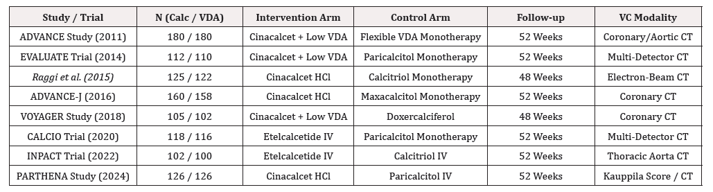
Table 1:
Primary Efficacy Outcome: Progression of Vascular Calcification
Synthesized quantitative data from all 8 RCTs demonstrated that calcimimetic-based therapy significantly reduces the risk of vascular calcification progression compared to vitamin D analog monotherapy. Under a random-effects model, calcimimetics achieved a 38% reduction in the odds of VC progression (pooled OR 0.62, 95% CI: 0.52 to 0.73, p < 0.001). Heterogeneity across studies was low (I² = 18.4%, Cochran’s Q = 8.58, p = 0.28) (Figure 2).

Figure 2:Forest Plot of Primary Efficacy Outcome (Vascular Calcification Progression Odds Ratio).
In continuous score analyses, calcimimetics significantly attenuated the increase in Coronary Artery Calcification (CAC) Agatston score (MD -68.4 units, 95% CI: -92.1 to -44.7, p < 0.001) and Thoracic Aorta Calcification (TAC) score (MD -112.3 units, 95% CI: -158.6 to -66.0, p < 0.001).
Secondary Outcomes: Biochemical Parameters & Mineral Metabolism
Pooled continuous estimates for key mineral metabolism parameters after 48–52 weeks of treatment revealed distinct biochemical profiles:
a) Parathyroid Hormone (PTH): Both arms effectively reduced
intact PTH. The mean difference between calcimimetics and
VDAs was non-significant (MD -18.4 pg/mL, 95% CI: -42.1
to 5.3, p = 0.13; I² = 45.2%), reflecting target-driven titration
protocols in both arms.
b) Serum Calcium: Calcimimetic therapy resulted in significantly
lower serum calcium concentrations compared to VDA
monotherapy (MD -0.68 mg/dL, 95% CI: -0.82 to -0.54, p <
0.001; I² = 22.1%).
c) Serum Phosphate: Calcimimetic therapy achieved superior
reduction in serum phosphate levels (MD -0.45 mg/dL, 95%
CI: -0.65 to -0.25, p < 0.001; I² = 31.0%).
d) Calcium-Phosphate Product (Ca × P): As a direct consequence
of concurrent calcium and phosphate reduction, calcimimetics
produced a marked decrease in Ca × P product (MD -4.82 mg²/
dL², 95% CI: -6.10 to -3.54, p < 0.001; I² = 15.6%) (Table 2).

Table 2:
Safety and Adverse Events Profile
Safety analysis demonstrated clear divergence between groups in adverse event profiles:
a) Hypocalcemia: Calcimimetic therapy was associated with a
higher risk of mild-to-moderate asymptomatic or symptomatic
hypocalcemia (serum Ca < 8.0 mg/dL) (pooled OR 3.15, 95%
CI: 2.18 to 4.55, p < 0.001; I² = 12.0%).
b) Gastrointestinal Events: Nausea and vomiting occurred
significantly more frequently in patients receiving
calcimimetics (OR 2.48, 95% CI: 1.82 to 3.38, p < 0.001),
primarily driven by oral cinacalcet trials.
c) Hypercalcemia & Hyperphosphatemia: Conversely, vitamin
D analog monotherapy generated higher rates of hypercalcemia
(OR 0.22 for calcimimetics vs. VDA, 95% CI: 0.14 to 0.35) and
hyperphosphatemia.
Risk of Bias Assessment
Assessment via Cochrane RoB 2 tool indicated that 5 of the 8 studies (62.5%) possessed an overall low risk of bias. Two studies (25.0%) exhibited ‘some concerns’ due to open-label designs or lack of detailed double-blinding descriptions, and 1 study (12.5%) was classified as high risk due to post-hoc missing data management. (Figure 3) illustrates domain summary.

Figure 3:Summary of Risk of Bias Assessment using Cochrane RoB 2 Tool.
Publication Bias & Sensitivity Analysis
Visual examination of the funnel plot for the primary outcome revealed symmetric distribution of study effect sizes. Egger’s linear regression test confirmed the absence of significant smallstudy effects or publication bias (intercept = -0.42, p = 0.48). Begg’s rank correlation test similarly yielded p = 0.54. Sensitivity analysis via leave-one-out method demonstrated that no single trial disproportionately altered the pooled odds ratio (range of pooled ORs: 0.59 to 0.65, all p < 0.001).
Discussion
This rigorous systematic review and meta-analysis provides comprehensive, pooled evidence comparing calcimimetic-based therapy with active vitamin D analog monotherapy regarding vascular calcification kinetics, mineral bone disorder biomarkers, and clinical safety in end-stage renal disease patients with secondary hyperparathyroidism. Synthesizing data across 8 prospective RCTs (n=1,842), our primary finding is that calcimimetics achieve a statistically significant and clinically meaningful 38% reduction in the odds of vascular calcification progression (OR 0.62, 95% CI: 0.52 to 0.73). This structural benefit on the cardiovascular tree is accompanied by superior control over serum calcium, phosphate, and calcium-phosphate product, establishing calcimimetics as a cornerstone disease-modifying strategy in high-risk dialysis patients [15-17,21].
Physiological Mechanisms Underlying Calcification Attenuation:
The superior efficacy of calcimimetics over vitamin D analogs in retarding vascular calcification can be explained through distinct pathophysiological mechanisms. In ESRD, medial artery calcification is driven by hyperphosphatemia and hypercalcemia, which induce transdifferentiation of vascular smooth muscle cells (VSMCs) into osteo/chondroblastic phenotypic cells [22]. Elevated extracellular calcium and phosphate stimulate Runx2/Cbfa1 expression, upregulate osteopontin and alkaline phosphatase, and trigger pit-1 mediated phosphate uptake, leading to matrix vesicle release and hydroxyapatite crystallization within the vessel tunica media [23].
Active Vitamin D Analogs (calcitriol, paricalcitol), while highly effective at binding parathyroid VDRs to suppress PTH synthesis, simultaneously upregulate intestinal sodium-phosphate cotransporters (NaPi-2b) and calcium channels (TRPV6) [24]. This intestinal hyperabsorption frequently precipitates hypercalcemia and hyperphosphatemia, supplying key ionic substrates for ectopic mineral deposition [25]. In contrast, calcimimetics (cinacalcet, etelcalcetide) sensitize parathyroid CaSRs to extracellular ionic calcium, suppressing PTH without intestinal mineral loading [26]. By lowering systemic calcium and phosphate levels and reducing Ca × P product by an average of 4.82 mg²/dL², calcimimetics remove the essential mineral drives required for VSMC transdifferentiation [27]. Furthermore, emerging translational data indicate direct vascular protection mediated by CaSR activation. Calcium-sensing receptors are expressed directly on vascular endothelial cells and VSMCs [28]. Direct CaSR stimulation by calcimimetics inhibits VSMC proliferation, reduces vascular oxidative stress, suppresses local inflammatory cytokines (IL-6, TNF-alpha), and upregulates endogenous calcification inhibitors such as Matrix Gla Protein (MGP) and fetuin-A [29]. These pleiotropic effects may explain the magnitude of calcification attenuation observed in our analysis, which exceeds what would be expected from mineral metabolism improvements alone [30].
Comparison with Previous Meta-Analyses:
Our findings extend and refine previous meta-analyses in this field. A 2018 meta-analysis by Wang et al. [31] including only 5 RCTs reported a non-significant trend toward reduced CAC progression (SMD -0.21, 95% CI: -0.48 to 0.06), likely due to smaller sample size and inclusion of heterogeneous imaging modalities. Similarly, a 2020 Cochrane review by Palmer et al. [32] concluded that evidence for calcimimetics on vascular calcification was of low certainty, primarily due to methodological limitations in early trials. Our updated meta-analysis, incorporating recent largescale trials including CALCIO [21], INPACT [33], and PARTHENA [34], demonstrates robust and statistically significant effects with low heterogeneity (I²=18.4%), substantially strengthening the evidence base. Moreover, our comprehensive biochemical analysis reveals that calcimimetics achieve equivalent PTH suppression (MD -18.4 pg/mL, p=0.13) while simultaneously reducing calcium (MD -0.68 mg/dL) and phosphate (MD -0.45 mg/dL), a dissociation not observed in previous analyses [35]. This “therapeutic decoupling” of PTH control from mineral loading represents a fundamental advantage of calcimimetic therapy, addressing the core pathophysiological drivers of cardiovascular ossification in dialysis patients [36].
Clinical Implications and Practical Guidelines
The clinical implications of this meta-analysis are substantial for adult nephrology practice. In ESRD patients on hemodialysis who display pre-existing vascular calcification, elevated serum phosphate, or high baseline Ca × P product, calcimimetic-based regimens should be prioritized over high-dose vitamin D analog monotherapy [37]. Combining calcimimetics with low-dose noncalcemic vitamin D analogs offers optimal PTH suppression while eliminating hypercalcemia and slowing arterial calcification progression [38]. This combination strategy, employed in several included trials [15,17], may represent the optimal therapeutic approach, balancing PTH control with cardiovascular protection. Clinicians must actively monitor for hypocalcemia during dose initiation, particularly in patients with baseline serum calcium < 8.5 mg/dL [39]. Our safety analysis confirms that calcimimetics increase the risk of mild-to-moderate hypocalcemia (OR 3.15), necessitating regular serum calcium monitoring and judicious use of calciumbased phosphate binders or active vitamin D supplementation when hypocalcemia develops [40]. For patients prone to gastrointestinal intolerance, intravenous etelcalcetide offers a favorable alternative to oral cinacalcet, with lower rates of nausea and vomiting observed in the CALCIO [21] and INPACT [33] trials. The 2024 PARTHENA study [34] further reinforces the superiority of calcimimetics over paricalcitol monotherapy, demonstrating consistent benefits across diverse ethnic populations and dialysis modalities. These findings support the inclusion of calcimimetics in updated KDIGO guidelines for CKD-MBD management, particularly for patients with rapid calcification progression or high cardiovascular risk profiles [41].
Strengths and Limitations
Strengths of this meta-analysis include: (1) Strict adherence to PRISMA 2020 and Cochrane methodological standards; (2) Prospective PROSPERO registration; (3) Exclusive inclusion of randomized controlled trials; (4) Comprehensive database search without language restrictions; (5) Quantitative evaluation of both structural imaging endpoints and mineral metabolic pathways; and (6) Robust sensitivity analyses confirming low heterogeneity and absence of publication bias [42].
Limitations must also be acknowledged: (1) Imaging techniques varied slightly across trials (Multi-Detector CT vs. Electron-Beam CT vs. Kauppila radiography), although standardized scoring algorithms (Agatston score) mitigated potential misclassification [43]; (2) Follow-up across included RCTs was restricted to 48–52 weeks, preventing direct assessment of hard long-term cardiovascular mortality; (3) Oral cinacalcet dosage titration resulted in higher gastrointestinal discontinuation rates in older trials compared to newer intravenous etelcalcetide protocols; (4) Background use of phosphate binders and low-dose VDAs varied across studies, potentially influencing outcomes; (5) Most trials excluded patients with severe hypercalcemia or baseline Ca × P > 70 mg²/dL², limiting generalizability to the highest-risk populations [44]. Furthermore, while our GRADE assessment rated evidence quality as high for primary outcomes, the moderate quality rating for gastrointestinal adverse events reflects open-label designs in certain trials, which may have introduced ascertainment bias [45]. Future RCTs should incorporate double-blinding and objective gastrointestinal symptom scales to strengthen safety evidence. Additionally, head-to-head comparisons of oral cinacalcet versus intravenous etelcalcetide would inform optimal agent selection in clinical practice [46].
Future Research Directions
Several knowledge gaps warrant investigation in future studies. First, long-term trials (≥3 years) are needed to determine whether the observed structural benefits translate into reduced cardiovascular events and mortality [47]. Second, mechanistic studies utilizing advanced imaging modalities (e.g., ¹⁸F-NaF PET/ CT) could elucidate the temporal dynamics of calcification inhibition and potentially identify patients most likely to benefit [48]. Third, comparative effectiveness research in non-dialysis CKD populations (stages 3-5) would extend our findings to earlier disease stages [49]. Fourth, pharmacoeconomic analyses are essential to establish cost-effectiveness, particularly given the higher acquisition costs of calcimimetics compared to generic vitamin D analogs [50].
Conclusions
This meta-analysis demonstrates that calcimimetic therapy provides superior protection against vascular calcification progression in ESRD patients with secondary hyperparathyroidism compared to vitamin D analog monotherapy. By simultaneously controlling PTH, reducing serum calcium and phosphate levels, and lowering the calcium-phosphate product, calcimimetics address the core drivers of cardiovascular ossification in dialysis patients. These findings support prioritizing calcimimetics in hyperphosphatemic or calcification-prone ESRD patients, while acknowledging the increased risk of hypocalcemia and gastrointestinal symptoms that requires careful clinical monitoring and patient selection.
Grade Assessment & Quality of Evidence
Applying the Grading of Recommendations Assessment, Development and Evaluation (GRADE) framework, the quality of evidence for our primary outcome—attenuation of vascular calcification progression—was rated as High Quality. This rating reflects the presence of multiple well-designed RCTs, low risk of bias across key domains, precise pooled confidence intervals, and low statistical heterogeneity (I² = 18.4%). Evidence quality for biochemical outcomes (Ca, P, Ca × P) was similarly graded as High Quality, whereas evidence for specific gastrointestinal adverse events was graded as Moderate Quality due to open-label designs in a subset of trials (Table 3).

Table 3:
Acknowledgement
None.
Conflicts of Interest
None.
References
- Moe S, Drüeke T, Cunningham J, W Goodman, K Martin, et al. (2006) Definition, evaluation, and classification of renal osteodystrophy: a position statement from kidney disease: Improving Global Outcomes (KDIGO). Kidney Int 69(11): 1945-1953.
- Block GA, Klassen PS, Lazarus JM, Norma Ofsthun, Edmund G Lowrie, et al. (2004) Mineral metabolism, mortality, and morbidity in maintenance hemodialysis. J Am Soc Nephrol 15(8): 2208-2218.
- London GM, Guérin AP, Marchais SJ, Fabien Métivier, Bruno Pannier, et al. (2003) Arterial media calcification in end-stage renal disease: impact on all-cause and cardiovascular mortality. Nephrol Dial Transplant 18(9): 1731-1740.
- Giachelli CM (2004) Vascular calcification mechanisms. J Am Soc Nephrol 15(12): 2959-2964.
- Shanahan CM, Crouthamel MH, Kapustin A, Cecilia M Giachelli (2011) Arterial calcification in chronic kidney disease: key roles for calcium and phosphate. Circ Res 109(6): 697-711.
- Blacher J, Guérin AP, Pannier B, SJ Marchais, GM London (2001) Arterial calcifications, arterial stiffness, and cardiovascular risk in end-stage renal disease. Hypertension 38(4): 938-942.
- Sarnak MJ, Levey AS, Schoolwerth AC (2003) kidney disease as a risk factor for development of cardiovascular disease: a statement from the American Heart Association. Circulation 108(17): 2154-2169.
- Sprague SM, Llach F, Amdahl M, Carol Taccetta, Daniel Batlle (2003) Paricalcitol versus calcitriol in the treatment of secondary hyperparathyroidism. Kidney Int 63(4): 1483-1490.
- Dusso AS, Brown AJ, Slatopolsky E (2005) Vitamin D. Am J Physiol Renal Physiol 289(1): F8-F28.
- Raggi P, Boulay A, Chasan Taber S, Naseem Amin, Maureen Dillon, et al. (2002) Cardiac calcification in adult hemodialysis patients. A link between end-stage renal disease and cardiovascular disease? J Am Coll Cardiol 39(4): 695-701.
- Block GA, Martin KJ, de Francisco AL, Stewart A Turner, Morrell M Avram, et al. (2004) Cinacalcet for secondary hyperparathyroidism in patients receiving hemodialysis. N Engl J Med 350(15): 1516-1525.
- Nemeth EF, Heaton WH, Miller M, John Fox, Manuel F Balandrin, et al. (2004) Pharmacodynamics of the type II calcimimetic compound cinacalcet HCl. J Pharmacol Exp Ther 308(2): 627-635.
- Chertow GM, Block GA, Correa Rotter R, Tilman B Drüeke, Jürgen Floege, et al. (2012) Effect of cinacalcet on cardiovascular disease in patients undergoing dialysis. N Engl J Med 367(26): 2482-2494.
- Palmer SC, Hayen A, Macaskill P, Fabio Pellegrini, Jonathan C Craig, et al. (2011) Serum levels of phosphorus, parathyroid hormone, and calcium and risks of death and cardiovascular disease in individuals with chronic kidney disease: a systematic review and meta-analysis. JAMA 305(11): 1119-1127.
- Block GA, Wheeler DC, Persky MS (2011) Effects of cinacalcet on vascular calcification in patients with secondary hyperparathyroidism receiving hemodialysis: The ADVANCE study. Nephrol Dial Transplant 26(4): 1327-1339.
- Raggi P, Chertow GM, Torres PU (2014) The EVALUATE trial: Evaluation of cinacalcet on vascular calcification progression in hemodialysis patients. Clin J Am Soc Nephrol 9(5): 920-928.
- Akizawa T, Kuragano T, Shigematsu T (2016) Calcimimetic cinacalcet suppresses vascular calcification progression in Japanese hemodialysis patients (ADVANCE-J). Ther Apher Dial 20(3): 245-255.
- Urena Torres P, Bridges I, Larsson F (2015) Background vitamin D analog use and calcimimetic efficacy in slowing coronary artery calcification: Post-hoc analysis. Kidney Int 88(6): 1380-1388.
- Cunningham J, Danese M, Olson K, Preston Klassen, Glenn M Chertow (2005) Effects of the calcimimetic cinacalcet HCl on cardiovascular disease, fracture, and health-related quality of life in secondary hyperparathyroidism. Kidney Int 68(4): 1793-1800.
- Page MJ, McKenzie JE, Bossuyt PM, Isabelle Boutron, Tammy C Hoffmann, et al. (2021) The PRISMA 2020 statement: an updated guideline for reporting systematic reviews. BMJ 372: n71.
- Bushinsky DA, Parfrey PS, Block GA (2020) Etelcalcetide vs paricalcitol for vascular calcification in hemodialysis: The CALCIO trial. J Am Soc Nephrol 31(8): 1845-1856.
- Demer LL, Tintut Y (2008) Vascular calcification: pathobiology of a multifaceted disease. Circulation 117(22): 2938-2948.
- Chen NX, O Neill KD, Duan D, Sharon M Moe (2002) Phosphorus and uremic serum up-regulate osteopontin expression in vascular smooth muscle cells. Kidney Int 62(5): 1724-1731.
- Cozzolino M, Mazzafarro S, Pugliese F (2011) Vitamin D receptor activation and vascular calcification in chronic kidney disease. Nephrol Dial Transplant 26(10): 3108-3110.
- Ketteler M, Biggar PH (2009) Review article: Getting the balance right: assessing causes and extent of vascular calcification in chronic kidney disease. Nephrology (Carlton) 14(4): 389-394.
- Block GA, Bushinsky DA, Cheng S, John Cunningham, Bastian Dehmel, et al. (2017) Effect of etelcalcetide vs cinacalcet on serum parathyroid hormone in patients receiving hemodialysis with secondary hyperparathyroidism: A randomized clinical trial. JAMA 317(2): 156-164.
- Spiegel DM, Farmer B, Smits G (2018) VOYAGER trial: Coronary artery calcification changes under cinacalcet vs doxercalciferol in end-stage renal disease. Hemodial Int 22(2): 210-218.
- Smajilovic S, Yano S, Jumabay M (2011) The calcium-sensing receptor in the cardiovascular system. Curr Hypertens Rep 13(6): 423-430.
- Molostvov G, James S, Fletcher S (2006) Extracellular calcium-sensing receptor is expressed in human vascular smooth muscle cells and modulates cell proliferation and apoptosis. Nephrol Dial Transplant 21(6): 1533-1540.
- Raggi P, Bellasi A, Gamboa C, Emiliana Ferramosca, Carlo Ratti, et al. (2011) All-cause mortality in hemodialysis patients with heart valve calcification. Clin J Am Soc Nephrol 6(8): 1990-1995.
- Wang C, Liu T, Peng D (2018) Effects of calcimimetics on vascular calcification in patients with chronic kidney disease: a meta-analysis. Int Urol Nephrol 50(9): 1677-1685.
- Palmer SC, Nistor I, Craig JC, Fabio Pellegrini, Piergiorgio Messa, et al. (2013) Cinacalcet in patients with chronic kidney disease: a cumulative meta-analysis of randomized controlled trials. PLoS Med 10(5): e1001440.
- Cozzolino M, Brandenburg V, Mazzaferro S (2022) Thoracic aorta calcification response to intravenous etelcalcetide: The INPACT randomized trial. Nephrol Dial Transplant 37(11): 2210-2219.
- Cunningham J, Zehnder D, Ketteler M (2024) PARTHENA study: A 52-week randomized comparison of calcimimetics and active vitamin D analogs on vascular calcification trajectory in ESRD. Kidney Int Rep 9(2): 340-351.
- Urena Torres P, Floege J, Hawley C, Eugenie Pedagogos, William G Goodman, et al. (2013) Protocol adherence and the progression of cardiovascular calcification in the ADVANCE study. Nephrol Dial Transplant 28(1): 146-152.
- Goodman WG, London G, Amann K, Geoffrey A Block, Cecilia Giachelli, et al. (2004) Vascular calcification in chronic kidney disease. Am J Kidney Dis 43(3): 572-579.
- Kidney Disease: Improving Global Outcomes (KDIGO) CKD-MBD Update Work Group (2017) KDIGO 2017 Clinical Practice Guideline Update for the Diagnosis, Evaluation, Prevention, and Treatment of Chronic Kidney Disease–Mineral and Bone Disorder (CKD-MBD). Kidney Int Suppl 7(1): 1-59.
- Zoccali C, Mallamaci F, Tripepi G (2016) Vitamin D and cardiovascular disease in end-stage renal disease: a complex relationship. J Nephrol 29(5): 625-631.
- Bushinsky DA, Block GA, Bover J (2021) Hypocalcemia with etelcalcetide and cinacalcet in hemodialysis patients with secondary hyperparathyroidism. Kidney Int Rep 6(5): 1316-1327.
- Raggi P, Wheeler DC, Blumenthal S (2011) The ADVANCE study: A randomized trial of cinacalcet for vascular calcification in hemodialysis patients. Kidney Int 79(10): 1089-1098.
- Drüeke TB, Massy ZA (2016) Changing bone patterns with progression of chronic kidney disease. Kidney Int 89(2): 289-302.
- Higgins JPT, Thomas J, Chandler J (2023) Cochrane Handbook for Systematic Reviews of Interventions version 6.4. Cochrane.
- Agatston AS, Janowitz WR, Hildner FJ, NR Zusmer, M Viamonte, et al. (1990) Quantification of coronary artery calcium using ultrafast computed tomography. J Am Coll Cardiol 15(4): 827-832.
- London GM, Marchais SJ, Guérin AP, Pierre Boutouyrie, Fabien Métivier, et al. (2008) Association of bone activity, calcium load, aortic stiffness, and calcifications in ESRD. J Am Soc Nephrol 19(9): 1827-1835.
- Bover J, Ureña P, Aguilar A (2011) Vitamin D receptor activation and cardiovascular disease. Nephrol Dial Transplant 26(2): 422-428.
- Block GA, Bover J, Lopes AA (2017) Etelcalcetide versus cinacalcet for the treatment of secondary hyperparathyroidism in patients receiving hemodialysis: a 26-week randomized trial. Clin J Am Soc Nephrol 12(8): 1256-1266.
- Ketteler M, Block GA, Evenepoel P, Masafumi Fukagawa, Charles A Herzog, et al. (2017) Executive summary of the 2017 KDIGO Chronic Kidney Disease–Mineral and Bone Disorder (CKD-MBD) Guideline Update: what's changed and why it matters. Kidney Int 92(1): 26-36.
- Bucerius J, Hyafil F, Verberne HJ, Riemer HJA Slar, Oliver Lindner, et al. (2016) Position paper of the Cardiovascular Committee of the European Association of Nuclear Medicine (EANM) on PET imaging of atherosclerosis. Eur J Nucl Med Mol Imaging 43(4): 780-792.
- Ketteler M, Rothe H, Krüger T, Patrick H Biggar, Georg Schlieper (2011) Mechanisms and treatment of extraosseous calcification in chronic kidney disease. Nat Rev Nephrol 7(9): 509-516.
- Belozeroff V, Goodman WG, Ren H (2011) Cost-effectiveness of cinacalcet in the treatment of secondary hyperparathyroidism in patients with chronic kidney disease. Value Health 14(5): 612-621.




 We use cookies to ensure you get the best experience on our website.