Case report 
 Creative Commons, CC-BY
Primary Neuroendocrine Carcinoma of the Cervix: A Case Report and Review of the Literature
*Corresponding author: Boujarnija Raihana, Hassan II hospital university, Fez, Morocco.
Received: December 03, 2019; Published: December 16, 2019
DOI: 10.34297/AJBSR.2019.06.001069
Abstract
Neuroendocrine carcinomas of the cervix are rare, and account for only 2% of all cervical cancers. the diagnostic and therapeutic management of these tumors is challenging and is extrapolated from other locations of neuroendocrine tumors. Rare cases of neuroendocrine tumors of the cervix have been reported in the literature and were associated with aggressive behavior and poor prognosis. We report the case of a 62 years old woman, diagnosed with metastatic cervical neuroendocrine carcinoma who received palliative chemotherapy with good control of her disease.
Keywords: Neuroendocrine carcinoma; immunohistochemistry; Chemotherapy
Introduction
Neuroendocrine tumors (NETs) are neuroendocrine cellderived malignancies that can occur in various locations. They are divided into four categories: small cell, large cell, atypical cell, and classical carcinoid tumors [1] . Neuroendocrine carcinoma of the cervix is a rare malignancy and accounts less than 2% of all cervical cancers [2] . In literature, there are very few reported studies and they involved only small series and case reports of neuroendocrine small cell cervical carcinoma. Furthermore, the patients are at advanced stages in most of the reported cases. It is difficult to manage these tumors. They are often diagnosed at an advanced stage and the prognosis is generally poor[2, 3]. The most common treatment for patients with advanced disease is chemotherapy based on etoposide and cisplatin. Through this case, we report our experience in the management of this particular tumor and discuss it in comparison with literature.
Case report
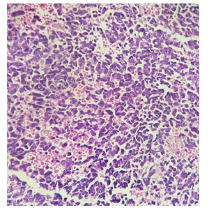
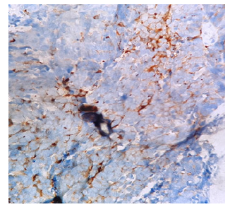
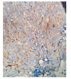
We report the case of a 62-years-old woman with a medical history of diabetes under oral antidiabetic drugs. She reported in July 2018 o occurrence of spontaneous low abundance metrorrhagia. The clinical examination revealed a large cervical hemorrhagic mass at the level of the cervix measuring 6cm. Cervical biopsy revealed an infiltrating malignant process and the immunohistochemical study showed a positive staining for the P16, synaptophysin (Figure 1), chromogranin A (+), and CD5/6. Thus, the cyto-architectural and immunohistochemical aspect was compatible with small cell carcinoma of the cervix () thoraco pelvic abdominal CT scan revealed a locally advanced cervical tumor with metastatic lymph nodes in addition to pulmonary and bone metastases (Figure 4).
The patient received first line chemotherapy based on cisplatin 80 mg / m2 and etoposide 100mg / m2 (j1, j2, j3) with bone modulating agents based on denosumab because of the presence of bone metastases. The patient received 3 cycles with partial response then continued until a total of 6 cycles. The evaluation have shown disease stability. Therefore, a follow-up of patient was decided. After 18 months of follow-up the patient is still with good clinical and radiological control (Figure 1)(Figure 2)(Figure 3)(Figure 4).
Discussion
Neuroendocrine tumors originate from Kulchitsky cells which can be found in all parts of the body, cervical localization is rare and represents only 3% of cervical tumors which are predominantly squamous cell carcinomas[4]. During the last two decades, and unlike squamous cell carcinoma of the cervix, an increase in the incidence of small cell neuroendocrine carcinomas has been observed. These tumors occur at a median age of 40 years (20-87) [5], which appears younger than for squamous cell carcinoma of the cervix. The clinical symptomatology is nonspecific it is most often metrorrhagia, recurrent leucorrhea or pelvic mass. The diagnosis is based on the histological study, neuroendocrine differentiation can be shown by many methods. The most important of all these methods is immunohistochemical study with chromogranin A, neuron specific enolase (NSE) which was implemented in this study. Chromogranin A is a more specific stain on this issue.
In the literature, there are studies that have been conducted to show the presence and significance of neuroendocrine differentiation in a specific tumor, especially on gastrointestinal tract and lungs, and these studies have concluded that this differentiation is related with poor prognosis and survival [6].Race, age and stage of the tumor seem to be the prognostic factors also smoking and advanced stage are reported to be poor prognostic factors for survival in patients with NE small cell carcinoma of the cervix . Of all these, the most important prognostic factor is lymph node metastasis. While the lymph node invasion in neuroendocrine tumors is more than 30% [7]. Given the strong trend towards regional and remote dissemination, the assessment should include abdomino-pelvic imaging, preferably magnetic resonance imaging. In order to improve ganglion staging FDG-PET is significantly more accurate than computed tomography (CT) and is recommended for loco-regional lymph node and extra pelvic staging. The metabolic dimension of the technique provides additional prognostic information, allowing a good follow-up of the target lesions, it becomes the tool of choice when one wishes to appreciate at best the effectiveness of a treatment [7].
The treatment of cervical neuroendocrine carcinomas is modeled after squamous cell carcinoma, taking into account the characteristics of neuroendocrine tumors of the lung. In the case of metastatic disease, palliative chemotherapy with cisplatin and etoposide or VAC- type chemotherapy (vincristine, adriamycin and cyclophosphamide) are indicated [8, 9] However, in the presence of small series and a few case reports it is difficult to analyze the effects of a treatment. Our patient was diagnosed at an advanced stage and having benefited from the systematic chemotherapy. After 18 months of follow-up the patient is still with good clinical and radiological control.
Conclusion
The cervix is a rare location of this subtype making the diagnosis very challenging and the confirmation is obtained by histology and immunohistochemistry. Conventional chemotherapy based on platinum and etoposide is still the mainstay of treatment in metastatic setting. More advanced research is required to better understand these tumors and allow development of effective drugs for this rare disease and improve the outcome of patients.
References
- Wells M, Nesland JM, Oster AG (2003) Epithelial tumors. In: Tavassoli FA, Devilee P, editors. World Health Organization classification of tumours: pathology and genetics of tumours of the breast and female genital organs. Lyon (France): IARC. Press 2003: 279.
- Pazdur R, Boromi P, Slayton R, Gould VE, Miller A, et al. (1981) Neuroendocrine carcinoma of the cervix: implications for staging and therapy. Gynecol Oncol 12(1): 120-128.
- Wang KL, Chang TC, Jung SM, Chen CH, Cheng YM, et al. (2012) Primary treatment and prognostic factors of small cell neuroendocrine carcinoma of the uterine cervix: a Taiwanese Gynecologic Oncology Group study. Eur J Cancer 48(10): 1484-1494.
- McCusker ME, Coté TR, Clegg LX, Tavassoli FJ (2003) Endocrine tumors of the uterine cervix: incidence, demographics, and survival with comparison to squamous cell carcinoma. Gynecol Oncol 88(3): 333-339.
- Cohen JG, Kapp DS, Shin JY, Urban R, Sherman AE, et al. (1999) Small cell carcinoma of the cervix: treatment and survival outcomes of 188 patients. AmJ Obstet Gynecol 72(1): 3-9.
- Piehl MR, Gould VE, Warren WH, Lee I, Radosevich JA, et al. (1988) Immunohistochemical identification of exocrine and neuroendocrine subsets of large cell lung carcinomas. Pathol Res Pract 183(6): 675-682.
- Wang KL, Chang TC, Jung SM, Chen CH, Cheng YM, et al. (2012) Primary treatment and prognostic factors of small cell neuroendocrine carcinoma of the uterine cervix: a Taiwanese Gynecologic Oncology Group study. Eur J Cancer 48(10): 1484-1494.
- Chan JK1, Loizzi V, Burger RA, Rutgers J, Monk BJ (2003) Prognostic factors in neuroendocrine small cell cervical carcinoma: a multivariate analysis. Cancer 97(3): 568-574.
- Hoskins PJ, Swenerton KD, Pike JA, Lim P, Aquino Parsons C, et al. (2003) Small-cell carcinoma of the cervix: fourteen years of experience at a single institution using a combined-modality regimen of involved-field irradiation and platinum-based combination chemotherapy. Clin Oncol 21(18): 3495-3501.


 We use cookies to ensure you get the best experience on our website.