
Mini-Review 
 Creative Commons, CC-BY
Carvedilol Improves Sorbitol Dehydrogenase and Heart Rate Variability Indices Thereby Ameliorating Cardiac Autonomic Neuropathy in T1DM Rats With Streptozotocin-Induced Diabetes
*Corresponding author: Akinlade Olawale Mathias, Cardiology unit, Internal Medicine Department, LAUTECH Teaching Hospital, Ogbomoso, Oyo State, Nigeria.
Received: February 25, 2020; Published: February 28, 2020
DOI: 10.34297/AJBSR.2020.07.001193
Abstract
Diabetic cardiac autonomic neuropathy (DCAN) is an important risk marker for all-cause and cardiac mortality, sudden death, and nephropathy progression [1] . The prevalence is very high among patients with diabetes Mellitus (DM). Moreover, despite intensive medical research on mechanism of DCAN and new medical treatments of patients living with this pathology, it still remains a major cause of mortality in diabetic patients and warrants further testing of novel therapeutic approaches. Carvedilol has been widely used in the treatment of heart failure, hypertension with or without DM for its effect on the autonomic system. Its role in the management of DCAN however has not been established. This study was designed to investigate the intervention of carvedilol in experimentally induced DCAN in rat model.
Methodology: DCAN was induced in 42 Wistar rats using streptozotocin (STZ) 50mg/kg. DCAN features were then assessed using both noninvasive time varying ECG and invasive biomarkers. The sham-fed control group received normal saline per oral while the experimental groups were treated with varying doses of Carvedilol.
Results: DCAN group had significantly (p<0.05) higher glucose (23.6±8.5 vs 4.68±0.91) and advanced glycated end products (63.54±2.09 vs 23.04±5.17) levels compared with controls. The level of antioxidants and sorbitol dehydrogenase (SD) (17.23±8.51 vs 32.94±5.07) activity were also reduced both of which were improved with administration of carvedilol. Furthermore, Heart Rate (HR) variability indices which were reduced with DCAN induction were also ameliorated with carvedilol. The HR variability indices correlated significantly with levels of biomarkers. This study shows that while SD activity and HR variability indices are deranged in DCAN, carvedilol may have a role in improving this indices and thus promising for the treatment of DCAN.
Keywords: Carvedilol; Diabetic Cardiac Autonomic Neuropathy; Diabetes mellitus; Heart rate variability; Sorbitol Dehydrogenase.
Background
Diabetic autonomic neuropathy (DAN) is one of the microvascular complication of diabetes with significant morbidity impacts. Diabetic cardiac autonomic neuropathy (DCAN) is one of the most studied and clinically important form of DAN: defined as the impairment of autonomic control of the cardiovascular system in patients with diabetes after exclusion of other causes [2] . The pathogenic mechanism is still uncertain and may be multifactorial [3] . Despite intensive study, no medication has been identified which can effectively stop or reverse DCAN once clinical features are apparent. Management so far is mainly based on prevention strategies which include intensive glycemic control, multifactorial intervention, lifestyle and dietary interventions.
The polyol pathway has been suggested to play an important role in the development of both neural and vascular complications in diabetes [4] . Inhibition of aldose reductase has been shown to reverse vascular and other complications in diabetics, while sorbitol dehydrogenase inhibition has been shown to exacerbate autonomic neuropathy in streptozotocin (STZ) induced diabetic rat model [2, 5, 6]. Carvedilol, a third generation β-adrenergic blocker had earlier been shown to improve survival outcomes in heart failure patients with autonomic neuropathy [7], but its role in DCAN had not been established. Although, some studies had shown its anti-oxidative functions in animal model with diabetes [8, 9, 10]. We investigate the effect of carvedilol on sorbitol dehydrogenase (polyol pathway enzyme) and resultant effect on cardiac autonomic neuropathy using non-invasive heart rate variability indices in STZinduced rat model.
Materials/Method
Animals
All experimental protocols were approved and done following the guiding principles of the University of Ilorin Ethical Review Committee (UERC). The research was approved by the same Ethical Review Committee (UERC) with approval no: UERC/ ASN/2019/1912. Male Wistar rats were used for the experiment. All animals were acclimatized to their environment for 2 weeks before commencement of the experiments. They were fed ad libitum and housed in pairs in wooden cages.
Experimental groups/Model development
Phase 1: Forty-two male Wistar rats were grouped into two; sham control group (n = 10) and
diabetic group (n =32). Type 1 DM was induced with high dose STZ at 50mg/Kg single dose under ketamine anaethesia. Rats with plasma glucose >16mmol/L on the third day after STZ-injection were randomized into different groups. Whole blood was taken from the proximal ventral tail vein for glucose measurement using a glucometer (Accu-Chek II Boehringer Mannheim Canada, Dorval, Quebec). Fasting plasma glucose levels, water intake and body weights were recorded at weekly intervals while other parameters were determined at the beginning and end of the experiment. Noninvasive holter measurement was also done before and after DM induction.
Phase2: This consists of the interventional phase which lasted for 28 days. Carvedilol was
administered in varying doses of 0.1mg/Kg, 1mg/Kg and 10mg/Kg respectively (DCAN 1A, 1B and 1C). A DCAN control group without carvedilol had only distilled water administered (DCAN 1D). The effects of DCAN on all the subgroups of the model were studied. These entail both non-invasive and invasive assessment of autonomic neuropathy. Time-varying, nonlinear and non-invasive methods were used to study cardiac autonomic dys-regulation from ECG records using holter ECG.
ECG and Measurement of time and frequency domain parameters
The Wistar rats were anesthesized with ketamine (75 mg/ kg) administered subcutaneously to stabilize the animal. The ECG signals were continuously recorded for at least 15 minutes in the limb, augmented and chest leads. Analysis was focused on the delineation technique using the ECG lead II, since it shows more amplitude P-, R-, S-, and T-waves. Lead II was achieved in the Wistar rat by placement of the positive electrode in the left xyphoid space while negative electrode was fixed around the right shoulder, in the same way as the Einthoven triangle (right arm position in the negative electrode and left leg position in the positive electrode).
Frequency and Time domain parameters including HR (mean heart rate), SDNN (standard deviation of normal-to-normal R-R intervals) and PNN50 (Percentage of successive RR intervals that differ by more than 50ms) were calculated from the HRV (Heart Rate Variability) data. The SDNN provided an estimate of overall HRV and sympathetic balance while the RMSSD yielded an estimate of the short term components of HRV and parasympathetic functions.
Invasive Assay
At the end of the intervention, 5-10 hours after the last dose of the medication, the rats were humanely euthanized after been anaesthetized, blood samples were collected via cardiac puncture after opening of the upper abdominal region. The samples were centrifuged at 1500 g x 15 minutes and the plasma samples micropipetted into plain bottles and immediately stored at a temperature of 0 -4oC. For the invasive assay, protein expression markers, which have been correlated with CAN and diabetes complications in previous studies and also recommended by the CAN subcommittee of the Toronto autonomic neuropathy working group, were assayed. These include; Nerve Growth Factor (NGF), Plasma Cathecholamines (CAT), Sorbitol Dehydrogenase (SD), Glutathione (GSH) and Total Anti-oxidant Capacity (TAC) [11, 12, [13, 14, 15]. These were then correlated with the time varying analysis.
Statistical Analysis
Data was analyzed using Statistical Package for Social Sciences (SPSS) software (Version 23.0; SPSS Inc, IL., USA) for windows. Results were expressed as the mean±SEM. Comparisons among groups was done using one-way ANOVA. Group means for two independent samples was compared using Student’s t-test and p values less than 0.05 was taken as statistically significant.
Results and Discussion
Diabetes induction
This study shows that STZ-induced diabetic rats developed significantly (p<0.05) higher glucose level compared with the controls (Figure 1)(Table 1). There was progressive weight loss in the DCAN group compared with the control group. The plasma levels of insulin and c-peptide were significantly (p<0.05) lower compared with the control. Carvedilol had no statistically significant effects on the levels of blood sugar, insulin and c-peptide. Diabetic induction was also seen to be associated with higher values of total cholesterol and triglyceride. Advanced Glycated Endproducts (AGEs) level showed a three-fold increase in the DCAN group compared with the control group. No significant effect was observed after carvedilol intervention in the level of AGEs (Table 1).
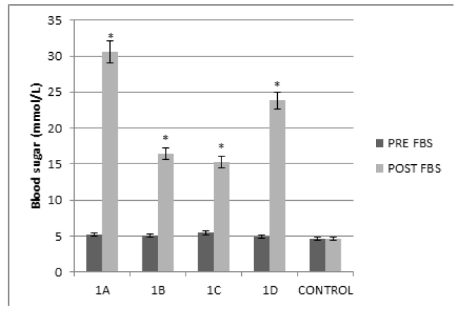
Figure 1: Showing Blood sugar before and after DM induction. Pre FBS=Fasting blood sugar before DM induction across each group as outlined in the methodology, Post FBS= Fasting blood sugar after DM induction, *p < 0.05 compared with normal control.
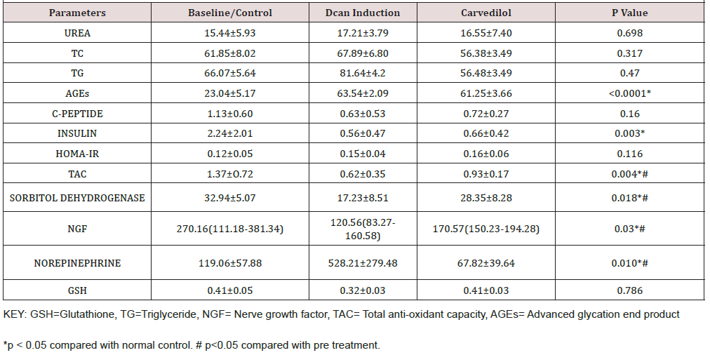
Table 1: Showing Laboratory Parameters And Overal Effect Of Dcan Induction And Carvedilol Administration On Serum Biochemical Parameters.
Effect of DCAN on sorbitol dehydrogenase, nerve growth factor and anti-oxidants
STZ-induced DCAN significantly reduces sorbitol dehydrogenase (SD) activity, alongside the anti-oxidants. SD is an enzyme in the polyol pathway which converts sorbitol to fructose. Studies have shown that sorbitol along with other metabolite mediates the neurotoxicity effect of chronic hyperglycaemia. Carvedilol however, increases the SD activity significantly (p<0.05) compared with the control (Figure 2). Similar trends were also observed for glutathione and total anti-oxidant capacity which were also improved with carvedilol administration (Figure 1)(Figure 2)(Table 1).
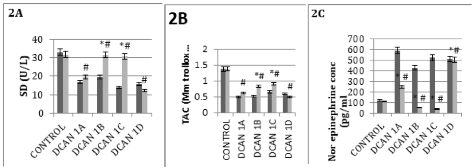
Figure 2: Comparing the effect of varying doses of carvedilol on significant laboratory parameters. Carvedilol at 1mg/Kg and 10mg/Kg significantly improves sorbitol dehydrogenase and total antioxidant capacity in (Figure 2) respectively while reducing norepinephrine concentration. (2C). *p < 0.05 compared with normal control, # p<0.05 compared with pre-treatment. (Deep color bar is preinterventrion, Light color bar is post intervention).
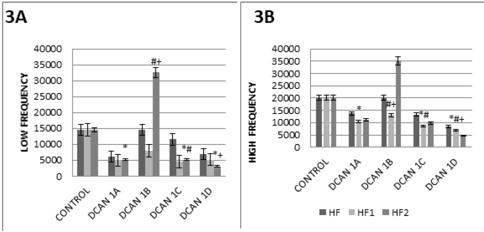
Figure 3: Showing Type 1 Dcan Frequency Domain 3A: The frequency domain was reduced after DM induction, but was significantly increased by administration of carvedilol (most significant effect was seen with 1mg/Kg dose) 3B: similar pattern as for low frequency was also seen with the high frequency domain LF=Low frequency, HF=High frequency. *p < 0.05 compared with normal control, # p<0.05 compared with DCAN control, +p<0.05 compared with pre treatment.
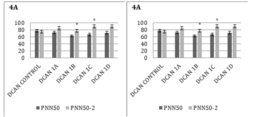
Figure 4: Showing Type 1 Dcan Time Domain SDNN index (SDNNI): Mean of the standard deviations of all the NN intervals for each 5 min segment of a 24 h HRV recording. PNN50: Percentage of successive RR intervals that differ by more than 50ms. *p < 0.05 compared with normal control. # p<0.05 compared with pre treatment.
The evaluation of autonomic activity using HR variability indices (frequency and time domain spectral) showed similar pattern with the biochemical markers. DCAN induction reduced both low and high frequency parameters. The group having 1mg/ Kg of Carvedilol had significantly (p<0.05) improved low and high frequency spectral activity. Most of the time domain parameters were not significantly altered with DCAN induction except for the triangular index, PNN50 and mildly the RMSSD. Carvedilol had no significant effect on the SDNN in the DCAN group (Figure 3)(Figure 4)
Discussion
The Sorbitol Pathway and oxidative stress in the Pathogenesis of Experimental Diabetic Autonomic Neuropathy
Numerous metabolic derangement associated with increased sorbitol pathway activity have been implicated in the pathogenic mechanism of diabetic complications. Earlier studies by Schmidt etal showed that inhibition of SD exacerbates autonomic neuropathy in rats with STZ-induced diabetes (5,6). This further supports the facts that sorbitol may plays a significant role in mediating nerve damage in diabetes autonomic neuropathy [16, 17]. Our study observed that while, STZ-induced DCAN was associated with reduced SD activity, an improvement in SD activity on exposure to carvedilol occurred which was further corroborated with the HR variability indices. A possible mechanism of production of reactive oxygen radicals through the sorbitol pathway is the transfer of electrons and proton ion from sorbitol to NAD+ faster than they can be utilized for synthesis of ATP and fatty acids [18, 19]. The antioxidant effect of carvedilol was seen in the significant improvement in TAC activity [20, 21]and GSH.
Heart rate variability indices in DCAN
This results shows that STZ-diabetic rats present changes in some of the heart rate variability indices especially the frequency domain spectral early in the course of diabetes. However, autonomic function evaluated by time-domain indices of HR variability was more pronounced about 28 days after STZ injection. Interestingly, all of these changes showed a negative correlation with plasma glucose and advanced glycated end products while positively correlated with SD and anti-oxidants markers. Earlier studies have suggested that DCAN have both parasympathetic and sympathetic dysfunction with early defective parasympathetic control, represented by persistent resting tachycardia and loss of beat-tobeat variation during deep respiration [22, 23, 24, 25]. Similar to what Faran et al. documented, we observed early changes in the PNN50 in the DCAN model while SDNN showed no significant changes [23]. SDNN had been shown to provide an estimate of overall HR variability and sympathetic balance while PNN50 correlates with overall parasympathetic events.
Our findings thus shows that parasympathetic dysfunction occur early in DCAN. Carvedilol had no effect on the SDNN but modest effect on improving the PNN50. This may be due to the fact that it takes longer time for the sympathetic dysfunction to manifest in abnormality of HR variability. Since, blood parameters shows significant increase in nor-epinephrine levels early after DCAN induction; it may then mean that HR variability indices lag behind plasma markers of sympathetic dysfunction. SDNN has important Our findings thus shows that parasympathetic dysfunction occur early in DCAN. Carvedilol had no effect on the SDNN but modest effect on improving the PNN50. This may be due to the fact that it takes longer time for the sympathetic dysfunction to manifest in abnormality of HR variability. Since, blood parameters shows significant increase in nor-epinephrine levels early after DCAN induction; it may then mean that HR variability indices lag behind plasma markers of sympathetic dysfunction. SDNN has important
Peripheral nerves have been shown to have structural changes which mimics those of human diabetic neuropathy and are observed to be preceded by hyperglycemia-induced biochemical abnormalities. Recent findings have emphasized the significance of vascular dysfunction, driven by metabolic impairments in the nerves, as an etiology of diabetic neuropathy. Non-enzymatic glycosylation of myelin components, deprivation of nerve growth factor, reduced endoneural blood flow, increased free oxygen radical activities and productions have also been hypothesized [35]. Our result strengthens the afore-mentioned showing impaired antioxidants activity and reduced NGF which correlates with the HR variability spectral events. On the other hand improved SD activity appears protective against the toxic neural effect in DCAN.
In conclusion, our results emphasis some well-known cardiac alterations using the STZ-diabetic rat model; such as increased AGEs, glucose and norepinephrine, with reduced SD activity, anti-oxidants and NGF, which all correlate with HR autonomic indices derangements. In agreement with previous researchers who showed that autonomic nerve structural abnormalities may not become apparent before functional changes, we believe that these changes could be due to early development of autonomic neuropathy in this model. The correlation observed between cardiovascular HR variability dysfunction and plasma biomarkers are in support of the possible presence of reversible neurological dysregulation.
References
- Yang B, Chon KH (2011) Assessment of cardiac autonomic neuropathy (CAN) in Type I diabetic mice. Conf Proc IEEE Eng Med Biol Soc 6560-6563.
- Verrotti A, Prezioso G, Scattoni R, Chiarelli F (2014) Autonomic Neuropathy in Diabetes Mellitus. Front Endocrinol (Lausanne). 1(5): 205.
- Lin Y-D, Hsu K-L, Wu E-T, Tsai M-S, Wang C-H, et al. (2008) Autonomic neuropathy precedes cardiovascular dysfunction in rats with diabetes. Eur J Clin Invest 38(9): 607-614.
- Steinmetz PR, Balko C, Gabbay KH (1973) The Sorbitol Pathway and the Complications of Diabetes. Vol. 288, New England Journal of Medicine 288(16): 831-836.
- Schmidt RE, Dorsey DA, Beaudet LN, Plurad SB, Williamson JR, et al. (1998) Effect of Sorbitol Dehydrogenase Inhibition on Experimental Diabetic Autonomic Neuropathy. J Neuropathol Exp Neurol 57(12):1175-1189.
- Schmidt RE, Dorsey DA, Beaudet LN, Plurad SB, Parvin CA, et al. (2001) Inhibition of Sorbitol Dehydrogenase Exacerbates Autonomic Neuropathy in Rats with Streptozotocin-Induced Diabetes. J Neuropathol Exp Neurol 60(12):1153-1169.
- Bristow MR, Gilbert EM, Abraham WT, Adams KF, Fowler MB, et al. (1196) Carvedilol produces dose-related improvements in left ventricular function and survival in subjects with chronic heart failure. Circulation 94(11): 2807-2816.
- Huang H, Shan J, Pan X-H, Wang H-P, Qian L-B, et al. (2007) Carvedilol improved diabetic rat cardiac function depending on antioxidant ability. Diabetes Res Clin Pract. 75(1): 7-13.
- Diogo C V, Deus CM, Lebiedzinska-Arciszewska M, Wojtala A, Wieckowski MR, et al. (2017) Carvedilol and antioxidant proteins in a type I diabetes animal model. Eur J Clin Invest 47(1): 19-29.
- Li X, Matta SM, Sullivan RD, Bahouth SW (2014) Carvedilol reverses cardiac insufficiency in AKAP5 knockout mice by normalizing the activities of calcineurin and CaMKII. Cardiovasc Res 104(2): 270-279.
- Bernardi L, Spallone V, Stevens M, Hilsted J, Frontoni S, et al. (2011) Methods of investigation for cardiac autonomic dysfunction in human research studies. Diabetes Metab Res Rev 27(7): 654-664.
- Aslam N, Kedar A, Nagarajarao HS, Reddy K, Rashed H, et al. (2015) Serum catecholamines and dysautonomia in diabetic gastroparesis and liver cirrhosis. Am J Med Sci 350(2): 81-86.
- Dimitropoulos G, Tahrani AA, Stevens MJ (2014) Cardiac autonomic neuropathy in patients with diabetes mellitus. World J Diabetes 5(1): 17-39.
- Porojan M, Costin S, Laura P, Anca C (2010) Autonomic neuropathy and plasma catecholamine in patients with diabetes mellitus | Request PDF. Rom J Intern Med 48(4): 341-345.
- Rolim LC, De Souza JST, Dib SA (2013) Tests for Early Diagnosis of Cardiovascular Autonomic Neuropathy: Critical Analysis and Relevance. Front Endocrinol (Lausanne) 4: 173.
- Bril V, Ono Y, Buchanan RA (2004) Sural Nerve Sorbitol in Patients with Diabetic Sensorimotor Polyneuropathy. Diabetes Care 27(5): 1160-1163.
- Yagihashi S, Mizukami H, Sugimoto K (2011) Mechanism of diabetic neuropathy: Where are we now and where to go? Vol. 2, Journal of Diabetes Investigation p. 18-32.
- Obrosova IG, Fathallah L, Lang HJ, Greene DA (1999) Evaluation of a sorbitol dehydrogenase inhibitor on diabetic peripheral nerve metabolism: A prevention study. Diabetologia 42(10): 1187-1194.
- Wolin MS (1996) Reactive Oxygen Species and Vascular Signal Transduction Mechanisms. Microcirculation 3(1): 1-17.
- Ayashi S, Assareh AR, Jalali MT, Olapour S, Yaghooti H (2016) Role of antioxidant property of carvedilol in mild to moderate hypertensive patients: A prospective open-label study. Indian J Pharmacol 48(4): 372-376.
- Dandona P, Ghanim H, Brooks DP (2007) Antioxidant activity of carvedilol in cardiovascular disease. J Hypertens 25(4): 731-741.
- Fazan RJ, Ballejo G, Salgado MCO, Moraes MFD, Salgado HC (1997) Heart Rate Variability and Baroreceptor Function in Chronic Diabetic Rats. Hypertension 30(3): 632-635.
- Ernst G (2017) Heart-Rate Variability-More than Heart Beats? Front Public Heal 5: 240.
- De Beck LD, Petersen SR, Jones KE, Stickland MK (2010) Heart rate variability and muscle sympathetic nerve activity response to acute stress: the effect of breathing. Am J Physiol Regul Integr Comp Physiol 299(1): R80-91.
- Young HA, Benton D (2018) Heart-rate variability: a biomarker to study the influence of nutrition on physiological and psychological health? Behav Pharmacol 29(2 and 3-Spec Issue): 140-151.
- Anichkov DA, Platonova AA (2009) Clinical Significance of Heart Rate Variability indexes derived from 5-Minute and 24-Hour ECG recordings in patients with Rheumatoid Arthritis. Rational Pharmacotherapy in Cardiol 5(1): 77-82.
- Fyfe-Johnson AL, Muller CJ, Alonso A, Folsom AR, Gottesman RF, et al. (2016) Heart Rate Variability and Incident Stroke. Stroke 47(6): 1452-1458.
- Mozaffarian D, Stein PK, Prineas RJ, Siscovick DS (2008) Dietary Fish and ω-3 Fatty Acid Consumption and Heart Rate Variability in US Adults. Circulation 117(9): 1130-1137.
- Poirier P (2014) Exercise, Heart Rate Variability, and Longevity. Circulation 129(21): 2085-2087.
- Soares-Miranda L, Sattelmair J, Chaves P, Duncan GE, Siscovick DS, et al. (2014) Physical Activity and Heart Rate Variability in Older Adults. Circulation 129(21): 2100-2110.
- Tsuji H, Larson MG, Venditti FJ, Manders ES, Evans JC, et al. (1996) Impact of Reduced Heart Rate Variability on Risk for Cardiac Events. Circulation 94(11): 2850-2855.
- Guzzetti S, Borroni E, Garbelli PE, Ceriani E, Bella P Della, et al. (2005) Symbolic Dynamics of Heart Rate Variability. Circulation 112(4): 465-470.
- Goldberger JJ, Challapalli S, Tung R, Parker MA, Kadish AH, et al. (2001) Relationship of Heart Rate Variability to Parasympathetic Effect. Circulation 103(15): 1977-1983.
- Sacha J (2014) Interplay between heart rate and its variability: a prognostic game. Front Physiol 5: 347.
- Lee PG, Hohman TC, Cai F, Regalia J, Helke CJ, et al. (2001) Streptozotocin-induced diabetes causes metabolic changes and alterations in neurotrophin content and retrograde transport in the cervical vagus nerve. Exp Neurol 170(1):149-161.




 We use cookies to ensure you get the best experience on our website.