Case Report 
 Creative Commons, CC-BY
Malignant Gastric Schwannoma: A Case Report & Review of Literature
*Corresponding author: Boujarnija raihana, Hassan II hospital university, Fez, Morocco
Received: January 09, 2020; Published: January 24, 2020
DOI: 10.34297/AJBSR.2020.07.001110
Abstract
Gastric schwannomas are unusual and account for only 0.2% of all gastric tumors. Given their rarity and the absence of randomized trials, the diagnostic and therapeutic burden of these tumors remains difficult. Like the latter, and despite multimodal treatment, their prognosis remains unfavorable. We report a new case of malignant gastric schwannoma and through the data of the literature we take stock of the different aspects of this rare entity.
Keywords: Malignant schwannoma; Gastric localization; Chemotherapy
Introduction
Schwannoma (Neurilimoma) Malignant (SWM) is also called malignant tumor of nerve cells, neurogenic sarcoma or neurofibrosarcoma. It is a tumor that develops from schwann cells (the sheath of the nerves) [1] . The schwannomas of the digestive tract remain rare and are mainly represented by gastric localizations [2, 3].
Gastric schwannoma accounts for only 0.2% of all gastric neoplasms. it is a benign tumor with an excellent prognosis after total resection [4] . Although the majority of schwannomas are benign, there are malignant forms, so-called malignant tumors of peripheral nerve sheaths which is the case of our patient [5, 6, 7, 8, 9, 10, 11]. We report the case of a 70 years old women with gastric malignant schwannoma and discuss the anatomopathological and evolutionary clinical features of this tumor entity.
Case Report
We report the case of a 70-years-old woman with a medical history of of hyperthyroidism under neo mercazol. The onset of clinical symptomatology dates back to three months before admission, with the appearance of an epigastric mass associated with intermittent pains without vomiting. The clinical examination revealed a mass at the level of the left hypochondrium, besides it did not show ascites or ganglion of Troisier. Fibroscopy revealed a large gastric mucosal tumor and several gastric biopsies were performed.
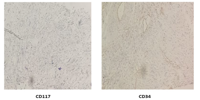
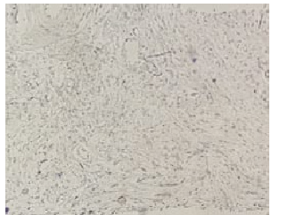
Biopsies performed at this level concluded fusiform cell proliferation with elongated nuclei, moderately irregular and hyperchromatic dense. Some nuclei are large, ovoid and sometimes nucleolate. no signs of adipocyte differentiation. Rare mitoses are observed focal. In some places the cells take on a more globular appearance with clear, non-vacuolar cytoplasm. Some lymphocyte elements dissociate this tumor proliferation. no signs of epithelial organization were seen. No foci of necrosis within these fragments. The Immunohistochemical study appears that the proliferation index is weak. Some nuclei are marked by ki67. Low and irregular nuclear expression of markers of liposarcomas: CDK4 and MDM2 no expression of Actin and H caldesmone muscle markers in the presence of internal controls, HMB 45 labeling was performed, it is negative. Tumor tumor cells do not express CD 117, CD34, Dog1 to rule out GIST or vascular tumor. SOX 10 is intensely expressed at the level of cell nuclei pointing to a schwann an origin. Before this aspect the pathological study concluded that the IHC profile favors a Schwannian conjunctive tumor.
Thoraco-abdominopelvic CT with contrast injection showed the presence of a well-limited tissue mass on the large gastric curvature measuring 33*66mm in diameter. intimate contact with the body of the pancreas without greasy separation border with the presence of two small nodular lesions in the seg VII and VI evoking secondary locations with peritoneal nodules measuring 7 mm in diameter for the largest. Our patient received palliative chemotherapy with doxorubicin with very good clinical tolerance and radiological stability (Figure 1) (Figure 2) (Figure 3) (Figure 4) .
Discussion
Gastric schwannoma is the most common among digestive schwannomas. But it represents only 0.2% of all gastric tumors, These tumors meet at all ages of life. But, usually occur between 40 to 60 years, with an average of 58 years. According to the series, we find a female preponderance [12, 13, 14, 15] as is the case of our patient, wich a sex-ratio equal 1 [16, 17, [18, 19, 20, 21, 22, [23, 24, 25]. The development of gastric schwannoma is low noise. It is often asymptomatic, and its discovery is most often fortuitous during a surgical procedure or a radiological or endoscopic examination [26]. In all the cases described in the literature, no specific symptomatology could be established to differentiate the SG from other gastric tumor lesions [23]. Other manifestations may be revealing: pyloric stenosis by pedicled tumor delivered by the pylorus, a palpable mass or distant metastasis.
The association of neurofibromatosis and schwannoma digestive is known without being mandatory. Thus in 1995 at the Avicenne Rabat Hospital (visceral surgical emergency department) a case was reported of a young patient aged 32, operated on a gastric malignant schwannoma, carrying many “café au lait” stains, with cutaneous tumors since birth, suggestive of VON RECKLINGHAUSEN [17]. In the presence of a schwannoma, as for a neurofibroma, it is necessary to look for the signs of an associated neurofibromatosis, in order to detect a familial susceptibility. The risk of cancer, including malignant schwannoma, would be higher in case of neurofibromatosis type 1 (NF 1) compared to the risk of the general population [18] the most classic CT appearance is that of a hypervascularized tumor with hypodense patches reflecting areas of necrosis or cystic degeneration [1,4].
The development of immunohistochemistry through the work of STERNBERGER in 1970 and TAYLOR in 1978 and the structural analysis allowed more identification on schwannoma among fusiform cell tumors of the application sometimes in question the initial diagnosis [11] Finally, the etiopathogenesis of this tumor remains very obscure, particularly in relation to Von Recklinghausen’s disease. The macroscopic appearance of gastric schwannomas presents itself as a firm, usually encapsulated tumor, well circumscribed, in the form of a multi-lobed, spherical or ovoid, developed along a nerve or eccentric, displacing the nerve fibers but never invading them, however this description “Is not specific [19, 20]. In the gastric wall: usually found in the submucosa or muscularis, which are repressed, dissociated by the tumor. Its base of implantation can be sessile or pediculated [19], The capsule can be missing in some cases. [15]. The size of the tumor varies between 0.5 and 15.5cm in diameter [21, 2]. At the cut, it appears, pearlywhite, yellowish or gray, even variegated in case of fatty infiltrate, necrotic or haemorrhagic zones [15].
Immunohistochemistry makes it possible to establish the diagnosis: the highly positive immunoreactivity to PS100 is compatible with a schwannoma (GISTs also express S100 in 10% of cases), other markers can be used. Nevertheless, the search for c-kit and CD34 is fundamental in order to eliminate GIST and, in the case of c-kit negativity, it is possible to carry out a specific mutation search for the kit and PDGFRA genes [6, 7, 9].
At immunohistochemistry schwannomas are still strongly positive for S100 protein. - Inconsistently Positive for: GFAP << Glial Fibrillary Acidic Protein >>, and Leu 7 >> Myelin Associated Glycoprotein>. -Really positive for CD34. -They are negative for: CD117 (or c-KIT), Desmin (DM), Smooth Muscle Actin (AML), Striated Muscle Actin (AMS), Myosin (MS).It is difficult to assert the benign or malignant nature of the tumor because mitosis, nuclear abnormalities and cellular polymorphism are not patho-gnomonic criteria. Malignant tumors can result from the degeneration of benign tumors or appear “novo” without precancerous lesions [8]. Sometimes only the evolution and the appearance of metastases make it possible to affirm the malignancy [10]. This was the case in our patient.
The elements suggesting malignancy are: the large size of the tumor, the break-in of the capsule, the adherence to neighboring organs, the calcifications and the necrotic haemorrhagic changes, the hypercellularity, the high mitotic activity of the appearance epithelioid and sometimes the diagnosis of malignancy is established only on the existence of metastases. The prognosis of gastric schwannomas is difficult to assess because of the exceptional nature or the scarcity of reported cases. According to STOUT [27] , malignant forms are always straight off. For others, it is the degeneration of a benign Schwannian tumor [25, 28, [29, 30, 31].
Complete surgical removal of the tumor is the gold standard. This removal can be difficult and sometimes incomplete due to intimate contact with large vessels or noble organs. Our patient was not able to benefit from a curative treatment due to the presence of several distant metastases. Remote metastases are localized to the lungs, liver and bones. They appear within an average of two years, which is shorter as long as there is an NF1 [32, 33, 21].
The first-line chemotherapy is based on anthracyclines. So far, it has not been shown that multidrug therapy is superior to doxorubicin monotherapy alone in overall survival. Therefore, chemotherapy with anthracyclines and ifosfamide may be the treatment of choice. MPNST responds poorly to radiotherapy and / or chemotherapy, however, this treatment must be undertaken [29, 30, 31]. The prognosis of gastric MPNST is poor and is associated with a rapid progression of the disease and a poor response to chemotherapy. Most patients die within 2 years, with an average 5-year survival rate of 23% [34] . In view of the reserved prognosis of these malignant tumors, surveillance of these patients is essential [24] .
Conclusion
Gastric schwannoma is one of the rare mesenchymal tumors. Its diagnosis is often late and difficult to establish preoperatively because of the absence of clinical, biological and radiological specificity of this tumor type, only the histopathological examination with immunohistochemical study allows establish the diagnosis of certainty.
The prognosis of mesenteric schwannomas is difficult to assess because of the exceptional nature or rarity of cases reported in the literature.
References
- Raber HM, Ziedses des Plantes CMP, Vink R, Klaase MJ (2010) Gastric schwannoma presenting as an incidentaloma on CT-scan andMRI. Gastroenterol Res 3(6): 276-280.
- Hong HS, Ha HK, Won HJ, Byun JH, Shin YM, et al. (2008) Gastric schwannomas: radiological features with endoscopic and pathological correlation. Clin Radiol 63(5): 536-542.
- Melvin WS, Wilkinson MG (1993) Gastric schwannoma: clinical and pathologic conditions. Am Surg 59(5): 293-296.
- Daimaru Y, Kido H, Hashimoto H, Enjoji M (1988) Benign schwannoma of the gastrointestinal tract: a clinicopathologic and immuno histochemical study. Human pathology 19(3): 257-264.
- Anandkumar A, Devaraj H (2013) Tumour immunomodulation: mucins in resistanceto initiation and maturation of immune response against tumours. Scand J Immunol 78(1): 1-7.
- Blay JY, Landi B, Bonvalot S, Monges G, Ray Coquard I, et al. (2005) Recommandations of management of GIST patients. Bull Cancer 92(10): 907.
- Geller DS, Gebhardt M (2006) Malignant peripheral nervesheath tumors (MPNST). Liddy Shriver Sarcoma Initiative.
- Serhane H (1989) Recklinghausen's disease and digestive schwannomas. Thèse Médicale pp. 67.
- Medeiros F, Corless C, Duensing A,Hornicq JL, Oliviera AN, et al. (2004) Kit-6negat if Gastrointestinal Stromal Tumors. AMJ Surg Pathol 28(7): 889-894.
- Jouvie J, Descottes B, Valleix D (1988) Tumeurs bénignes du duodé A propos de deux obs ervtions unschwannome et un léiomyome. J Chir 125(11): 646-649.
- Trojanowski JQ, Lee VM (1983) Monoclonal and polyclonal antibodies against neural antigens: diagnosis applications for studies of central and peripheral nervous system tumors. Hum Pathol 14(4): 281-285.
- Voltaggio L, Murray R, Lasota J, Miettinen M (2012) Gastric Schwannoma a Clinicopathologic study of 51 cases and critical review of literature. Hum. Pathol 43(5): 650-659.
- Hu J, Liu X, Ge N, Wang S, Guo J, et al. (2017) Role of endoscopic ultrasound and endoscopic resection for the treatment of gastric schwannoma. Medicine 96(25): 7175.
- Fujiwara S, Nakajima K, Nishida T, Takahashi T, Kurokawa Y, et al. (2013) Gastric schwannomas revisited: has precise preoperative diagnosis become feasible? Gastric Cancer 16: 318-323.
- Voltaggio L, Murray R, Lasota J, Miettinen M (2012) Gastric schwannoma: a clinicopathologic study of 51 cases and critical review of the literature. Human Pathology 43(5): 650-659.
- Handra Luca A, Nahon P, Flejou JF, Molas G, Dubois S, et al. (2001) Immunohistochemical and ultrastructural heterogeneity of digestive stromal tumors. Gastroenterol Clin Biol 25(6-7): 664-668.
- Amraoui M, Bougtab A, Alami H, Echarrab M, Louchi A, et al. (1995) Schwannome Malin Gastrique Au Cours De La Maladie De Recklinghausen : A Propos D’un Cas. Balafrej Médecine Du Maghreb 49 : 21-24.
- Leroy JP (1984) Sarcomas of nervous origin. Sem Hôp Paris 60: 2081-2086.
- Viros (1993) Denis Contribution to the study of gastric schwannoma: clinical, histological, radiological aspects, about a case. Thèse de médecine : Tours, France.
- Fayçal el Mrini (2005) Retroperitoneal Schwannoma: About 2 cases. Thesis Med.
- Bao guang Hu, Feng jie Wu, Jun Zhu, Xiao mei Li, Yu ming Li, et al. (2017) Gastric Schwannoma : A Tumor Must Be Included in Differential Diagnoses of Gastric Submucosal Tumors. Case Reports in Gastrointestinal Medicine pp. 8.
- Aminder Singh, Ankur Mittal, Bhavna Garg, Neena Sood Singh (2016) Schwannoma of the stomach: a case report. J Med Case Rep 10: 4.
- Vargas Flores E, Bevia Pérez F, Ramirez Mendoza P, Velázquez García JA, Ortega Román OA (2016) Laparoscopic resection of a gastric schwannoma: A case report. Int J Surg Case Rep 28: 335-339.
- Bouvier C, Daniel L, Figarella Branger D, Pellissier JF (1999) Tumors of the peripheral nervous system. Encycl Méd Chir 17-115-B-10.
- Dahl I (1977) Ancient neurilemmoma (schwannoma). Acta Pathol Microbiol Scand A 85(6): 812-818.
- Lin CS, Hsu HS, Tsai CH, Li WY, Huang MH (2004) Gastric schwannoma. J Chin Med Assoc 67: 583-586.
- Stout S (1935) The peripheral manifestation of the specific nerve sheat tumor (nerilemmoma). Am J Cancer 14: 751-796.
- Guivarch M (1987) Tumeurs rétropéritonéales, Rev Méd Interne 8(5): 493-502.
- Goaguen O, Boucher E, Pouit B, Soulard R, Lecharpentier M, et al. (2003) A reserved pronotic tumor: the melanocytic schwannoma. Neurochirurgie 49 : 31-38.
- Er U, Kazanci A, Eyriparmak T, Yigitkanli K, Senveli E (2007) Melanotic Schwannoma. Journal of Clinical Neuroscience 14: 676-678.
- Lyros O, Schickel S, Schierle K, Hoffmeister A, Gockel I (2017) Gastric schwannoma: rare differential diagnosis, acute upper gastrointestinal bleeding, gastric schwannoma: rare differential diagnosis of acute upper gastrointestinal (GI) bleeding. Z Gastroenterol 55(8): 761-765.
- Soualhi M, Elouazani H, Chaibainou A, Bouchantouf R (2004) Malignant tumor of the sheaths of the peripheral nerves, epithelioid type. Rev Pneumol Clin 60: 50-54.
- Topal O, Yilmaz T, Oğretmenoğlu O (2004) Giant malignant peripheral nerve sheath tumor of the neck in a patient with neurofibromatosis-1. Int J Pediatr Otorhinolaryngol 68(11) :1465-1467.
- Bees NR, Ng CS, Dicks Mireaux C, Kiely EM (1997) Gastric malignant schwannoma in a child. Br J Radiol 70: 952-955.


 We use cookies to ensure you get the best experience on our website.