Case report 
 Creative Commons, CC-BY
Transcatheter Aortic Valve Implantation: Transseptal Approach Using a Femoral Veno- Arterial Loop
*Corresponding author: Bertin Murielle, Department of Anaesthesia, Mediclinic City Hospital, Dubai Health Care City, building 37, Dubai, UAE.
Received: February 21, 2020; Published: February 26, 2020
DOI: 10.34297/AJBSR.2020.07.001175
Abstract
After successful introduction of transcatheter aortic valve implantation (TAVI) in 2002, TAVI has become an increasingly accepted therapy for the treatment of severe aortic stenosis in patients judged to be at high risk for open heart surgery. We describe anterograde transseptal access for TAVI in a patient where transfemoral access alone was impossible to achieve due to a heavily calcified stenotic aortic valve. It is an interesting and alternative approach to achieve TAVI.
Keywords: Transseptal, TAVI
Introduction
TAVI (transcatheter aortic valve implantation) is a less invasive procedure for aortic valve replacement. A crimped biological valve or self-expendable stent is inserted antegradely under fluoroscopy and deployed on a beating heart [1] . It is indicated for symptomatic severe aortic stenosis patients who are too fragile to have midline sternotomy and cardiopulmonary bypass. In our hospital TAVI has been done through transfemoral approach successfully for more than 60 patients. We describe a difficult case where the catheter couldn’t be passed through the aortic valve. Following transseptal puncure it was possible to pass a guide wire into the left ventricle and retrogradely through the aortic valve into the aorta. This helped the symptomatic patient who refused open heart surgery to have valve replacement. The outcome of replacement was very good. Some transseptal approach has been used in swine model and mitral valve replacement [2, 3].
Case Presentation
A 79-years-old man with symptomatic severe Aortic stenosis refused open-heart surgery but agreed to have TAVI. He has history of shortness of breath and recurrent pulmonary edema with repeated ICU admissions. He has history of smoking, and chronic obstructive pulmonary disease. Echocardiography preoperatively showed severe aortic stenosis, with a valve area of 0.7cm. and maximum gradient 85mm/Hg. Ejection fraction is 60 percent. He has no other valve disease. The arterial transfemoral approach TAVI was used first but it was not possible to pass the wire through aortic valve which was heavily calcified and stenotic.
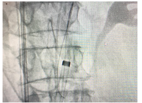
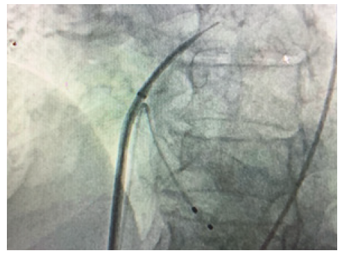
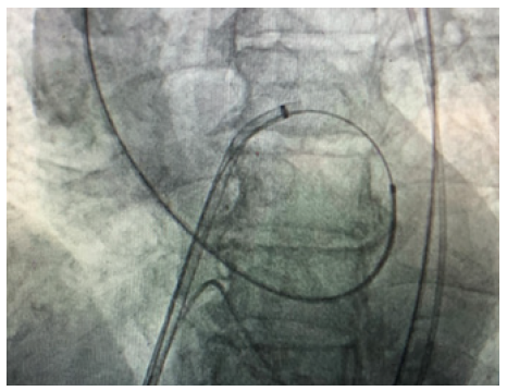
After repeated attempts it was decided to try an alternative approach. The right femoral vein was accessed and an 8F sheath inserted. Transeptal puncture was performed with a Brockenbrough needle and long Terumo wire passed through the left atrium to the left ventricle. The wire could then easily pass retrogradely through the aortic valve down to the descending aorta. The wire was then snared out of femoral artery. A multipurpose catheter was then passed over the wire and antegradely into the left ventricle and the guide wire exchanged. Finally, he nailed the wire to the right sheet of the TAVI. By pulling like this, the wire of the TAVI could cross the calcified valve. The cardiologist after passing the wire through aortic calcified valve put a balloon for the dilatation of the very stenotic valve. This venous-arterial wire loop is used to pass TAVI catheter through aortic valve. After the dilatation of the stenotic aortic valve with the balloon, the procedure continues as standard transfemoral approach. Medtronic Evolut valve was implanted successfully (Figure 1)(Figure 2)(Figure 3).

Figure 2: The wire goes from vena cava, right atrium,left atrium, left ventricle, aorta then femoral artery.
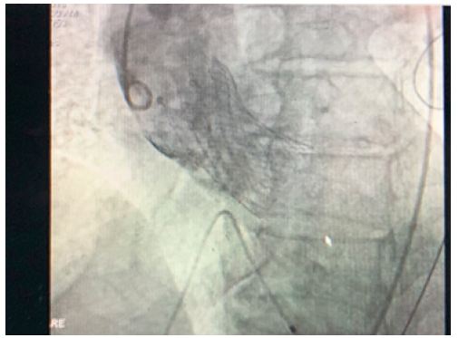
So direct implantation of Revolute R valve has been done, and the valve was perfectly in place. No need of post dilatation. It is a Medtronic Core Valve Evolut 34mm. The vascular femoral sheet is a proglide 6F (2mm). No major aortic regurgitation after the valve was in place and patient was in sinus rhythm. Temporary pacemaker fixed (Figure 4).
The first Echocardiography report just after the placement of the new valve shows a trivial paravalvular aortic regurgitation, concentric Left ventricle hypertrophy, type I Left Ventricle diastolic dysfunction, normal left ventricle function and trivial pericardial effusion. Patient shifted to ICU postop fully conscious, vital stable and without complaints. Discharged from hospital 3 days later without any complication. Two days after going home, patient came back to emergency room with palpitations and shortness of breath. Electrocardiogram shows rapid atrial fibrillation and patient was admitted in ICU with cordarone infusion. After one-hour patient converted to sinus rhythm.
Patient was sent home with Eliquis tablet and beta blocker tablet. Follow up of the patient after I month, 3 months and 6 months are normal with a well-functioning valve and sinus rhythm, stable, no complain after TAVI. The Echocardiography shows a well-functioning valve.
Discussion
TAVI is a less invasive treatment of the stenotic aortic valve avoiding sternotomy and cardiopulmonary bypass. A biological valve on self-expanding or balloon-expandable stent is inserted antegradely or retrogradely under fluoroscopy and deployed on the beating heart. Whether retrograde or anterograde TAVI should be considered the better approach is still an ongoing debate. Both techniques have been shown to be safe if performed by a dedicated multidiscipline team [4].
Anterograde access to the aortic valve is done through the apex of the left ventricle which need a left anterolateral minithoracotomy. Retrograde access to the aortic valve needs trans arterial access and is usually achieved transfemorally, or less frequent through the subclavian or carotid artery or the ascending aorta. The transfemoral artery TAVI can be done percutaneously, with less anesthesia requirement. The subclavian and transcarotid access are mostly done by a surgical cut-down. The transaortic access will need partial or full sternotomy or right partial thoracotomy. In our case here, patient was reluctant to have any surgery or cut down so an interesting advantage of using femoral vein and do a veno-arterial loop using a wire transseptally is an elegant way to perform TAVI percutaneously which didn’t require any surgery at all.
The femoral artery approach is considered as the most popular approach in our institute and more than 60 cases have been operated without any problem [5]. The Transseptal route has a significant value to access the mitral valve and has been used for the mitral valve replacement but non-published for aortic valve replacement.
In literature [4], different approaches of TAVI has been used and no data has shown superiority of one approach to another, so an individualized patient-cardiologist decision making process will be the most beneficial, taking advantage of the complementarity of different options. In our case as the catheter could not pass the calcified valve coming retrograde from the aorta, most probably transcarotid or subclavian access will show same problem with the obstructed valve. So, wire coming from femoral vein, vena cava, right atrium, going transseptal to left atrium, left ventricle then crossing the valve and going out to femoral artery. This wire nailed the catheter of TAVI and place it through the aortic valve inside the left ventricle. This shows an elegant way to avoid surgery and place the valve percutaneous. Dilatation of the valve with balloon straight after passing the wire open the valve, so classic TAVI can be performed after that [6].
Conclusion
In conclusion, this report describes the successful implantation of a Medtronic Core Valve Evolut in aortic position using a venous-arterial wire loop after establishment of transfemoral and transseptal access to left atrium then ventricle. The procedure is safe if the wire is place appropriate within the left ventricle then aorta. Thus, the venous transfemoral route might serve as a complementary access site in addition to transfemoral approach for TAVI. The all procedure can be done without cut down and percutaneously. This can be an advantage for patients who refuse any surgery or need less anesthesia.
Acknowledgement
We gratefully acknowledge Dr Taleb Majwal, interventional cardiologist Mediclinic City Hospital who perform this procedure, without which we could be able to do this case report.
Statement of Ethics
The study was done following the rules of our hospital ethic committee.
Disclosure Statement
The authors have no conflicts of interest to declare.
Funding Sources
None.
Author Contributions
All authors equally contributed.
References
- Sameer Arora, Jacob A Misenheimer, Radhakrishan Ramaraj (2017) Transcatheter Aportic Valve Replacement: Comprehensive Review and Present Status. Tex Heart Inst J 44(1): 29-38.
- Ulrich Schaefer, C Frerker, D Schewel, T Thielsen, F Meincke (2012) Transfemoral and Transseptal Valve-in-valve Implantation into a failing Mitral Xenograft with a Balloon-Expandable Biological Valve. Ann Thorac Surg 94(6): 2115-2118.
- Kenji Nakatsuma, N Saito, H watanade, B Bao, E Yamamoto, et al. (2017) Anterograde transcatheter aortic valve implantation using the looped Inoue Balloon technique: A pilot study in a swine model. J Cardiol 69(1) 260-263.
- S Bleiziffer, M Krane, MA Deutsch, Y Elhmidi, N Piazza, et al. (2013) Which way in? The necessity of multiple approaches to Transcatheter valve therapy. Curr Cardiol Rev 9(4): 268-273.
- Stephane Noble, Marco Roffi (2013) Overcoming the challenges of the transfemoral Approach in Transcatheter Aortic Valve Implantation. Interv Cardiol 8(2): 131-134.
- Pablo Salinas, R Taul Moreno, Jose Lopez-Sendon (2011) Transcatheter aortic valve implantation: Current status and future perspectives. World J Cardiol 3(6): 177-185.


 We use cookies to ensure you get the best experience on our website.