Case Report 
 Creative Commons, CC-BY
Femur Open Wedge Corrective Osteotomy and Gradual Deformity Correction
*Corresponding author: Charles Ang Poh Thean, International Islamic University Malaysia, Office of campus director, International islamic university malaysia, kuantan campus, Jalan Sultan Ahmad Shah, Bandar Indera Mahkota, 25200 Kuantan, Pahang, Malaysia.
Received: June 27, 2019; Published: July 05, 2019
DOI: 10.34297/AJBSR.2019.03.000713
Abstract
Deformity correction remains a challenge to treat. However, with the advancement of corrective osteotomy and illizarov external fixation, these complex deformities are better addressed. We report a case of atrophic non- union seen in a seventeen-year-old boy who was initially treated with locking plate following an open fracture to his left distal femur and tibial plateau. Infective causes was ruled out. Plain radiograph reveals a broken implant and atrophic non- union of his distal femur with thirty degrees medial angulation. He underwent removal of implant, corrective osteotomy and illizarov external fixation. At ten weeks post surgery, the deformity was completely corrected. The illizarov external fixator was removed at sixteen weeks post surgery, and he was able to ambulate without pain. Following a few sessions of physiotherapy over a period of three months, he was able to regain his knee full range of movement and was able to return to sports. Corrective osteotomy and illizarov external fixation remains the treatment of choice in chronic limb deformities, especially if suspicious of infection. This minimizes the risk of neurovascular tractional injury and implant related infection which will be disastrous to manage.
Keywords: Deformity correction, Open wedge osteotomy, Illizarov external fixation.
Case Report
A seventeen-year-old boy presented with pain and swelling of his left thigh following a futsal game. He denies any significant trauma during the game besides stomping on his left foot multiple times. He was still able to weight bear with pain after the game. Further history taking reveals that he was involved in a high energy motor vehicle accident on September 2017 and sustained open fracture supracondylar left femur and left tibial plateau. Open reduction and locking plate fixation were done for his femur and tibia at that point of time. He claims to be ambulating well with no pain, shortening or instability following the surgical intervention. He also denies any history of chronic discharge or fever.
Examination reveals a short limb and antalgic gait. Lateral bowing of his left femur along with 2cm shortening of his left femur. Deep tenderness over his distal femur and slight mobility appreciated over his distal femur. Otherwise there was no local signs suggestive of infection, such as increased warmth, erythema or sinus discharge.
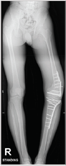
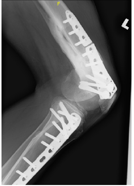
White blood cell count and C- reactive protein was not elevated with the reading of 3.8x10⁹/L and 0.5 mg/dL respectively. Erythrocyte sedimentation rate was slightly elevated at 30mm/hr. His lower limb radiograph reveals a broken implant and atrophic non- union of his distal femur with thirty degrees medial angulation (Figure 1). There was no significant antero- posterior angulation or translation (Figure 2). The radiograph was not suggestive of any long-standing infective process or osteomyelitis.

Figure 1: Full length antero- posterior plain radiograph reveals a broken implant, atrophic non- union of left distal femur with thirty degrees medial angulation.

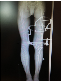
Figure 4:Full length antero- posterior plain radiograph at ten weeks post operative shows compete correction of the angular deformity
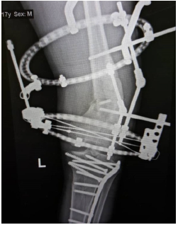
He underwent removal of implant, corrective osteotomy, left Illizarov external fixation and gradual deformity correction (Figure 3). Intra- operatively there was no local signs of infection to the distal femur and intra- operative cultures came back nil of growth/ organism. At ten weeks post-operative the deformity was corrected (Figure 4).
Discussion
Distal femur fractures show two peaks. It is seen in both young and old patients. In the young, it is usually a sequelae of a high energy road traffic accident and in the elderly from a trivial fall. Precise reduction and fixation of distal femur fractures with adequate stability allowing early mobilization is crucial. Nonunions of distal femur do not commonly occur. However, if it happens it causes significant morbidity and remains a nightmare to treat. The typical diagnostic criteria of non-union are pain and tenderness over the fracture site along with serial radiographic evidence showing no visible progressive signs of healing for three months, six months after the fracture [1]. In this case report, the fracture occurred twenty-six months ago and he still experienced pain and tenderness over the fracture site despite. We do not have a three months serial radiograph on him, however at twenty-six months following the fracture, a clear visible fracture line is still seen, indicating a non-union of his left distal femur.
Non-unions are broadly classified into septic and aseptic non-unions. Aseptic non-union is further divided into atrophic or hypertrophic. Atrophic non- union is avascular, nonviable and avital. It is associated with inadequate or poor vascularity with poor healing. Radiographically, it exhibits minimal callus formation filling the fracture gap surrounded by fibrous tissue. Hypertrophic non- union is said to be hyper vascular, viable and vital and occurs due to inadequate immobilization. The vascularity and healing is adequate. Radiographically, hypertrophic non- union shows increased callus formation in a horseshoe or elephant foot pattern [2]. As evident via clinical history and plain radiographs in this case, there was minimal callus seen around the fracture site indicating an atrophic non- union. The likely cause of this atrophic non- union is due to the high energy trauma he sustained leading to avascular, nonviable and avital tissues around the fracture site. Furthermore, the internal fixation he underwent further disrupt the blood supply over the fracture site contributing to the non-union.
Paul J Harwood et al. categorized the causes of non-union into four main groups, namely due to deficient of bone producing cells, deficient of signaling molecules, deficient of stability and deficient of bone conducting framework [3]. Craig S. Roberts et al. on the other hand categorized the causes of non-union into two main categories, namely the systemic causes and local causes. Systemic causes such as malnutrition, diabetes mellitus, cigarette smoking and nicotine use, osteoperosis and use of nonsteroidal anti- inflammatory drugs have been said to be the cause on non-union. As for the local causes, impaired vascularity, unstable fixation, presence of bone gap, infections, mal- alignment or rotation, lack of stimulation (eg: weight bearing), impact of injury (high- energy versus low- energy) and iatrogenic factors such as aggressive periosteal stripping plus local trauma to soft tissue and bone vascularity during fixation are the causes of non-union [4]. This patient does not have any significant systemic disorder contributing to his non-union. However, he has multiple contributing local causes such as impaired vascularity, high magnitude of injury and iatrogenic disruption to periosteum, bone and soft tissue during fixation.
Edward K. Rodriguez et al. in 2013 conducted a multi- centre, retrospective case control study on the predicting factors for non- union of distal femoral fracture following lateral locking plate fixation. He concluded that the only statically significant contributing factors to non- unions were compound fractures (open fractures), presence of an local infection, the use of a stainless steel implant and being obese with a body mass index of above 30 [5]. In this case, he sustained an compound fracture to his left distal femur
Deformity is defined as any deviation from the normal anatomy [6]. This includes any abnormalities of length, rotation, translation or angulation. This is assessed both clinically and radiographically. Clinically, first assess the frontal plane alignment with the patient standing straight.
Look for pelvic tilt, genu varum/ valgum, foot varus/ valgus/ abducrus/ adducrus/ supinated/ pronated, any obvious diaphyseal deformity and trendelenburg sign. Then assess the rotation alignment by looking at the patellar orientation and foot orientation. For length discrepancy, look for pelvic tilt, knee flexion or equinus stance, and the use of blocks for measurement. Then assess the sagittal plane alignment (standing lateral view) for spine lordorsis/ kyphosis, any hip flexion deformity, knee flexion or recurvatum deformity, ankle equinus or calcaneus, flat foot or cavus foot. Finally, assess the gait, patellar and foot progression angle. All this deformity is then confirmed clinically in sitting, supine and prone position. Radiographically, the normal anatomy is needed for comparison, in order to ascertain that the limb is abnormal and thereby deformed. Perform a long limb standing antero- posterior and lateral radiograph to evaluate the lower limbs. The anatomic axis is a line that bisects the medullary canal of the long bone longitudinally into two equal parts.
The mechanical axis of the lower limb is a point from the centre of the femoral head to the midpoint of the ankle. The normal mechanical axis deviation (MAD) is 1mm to 15mm medial to the center of the knee joint. MAD above 15mm medial to the knee midpoint indicates a varus malalignment and a MAD lateral to the knee midpoint indicates a valgus malalignment. This is known as the mechanical axis deviation test. Secondly run the malalignment test to determine the origin of the frontal plane malalignment. Draw the individual mechanical axis of the femur and tibia then measure the lateral distal femoral angle (LDFA) and medial proximal tibia angle (MPTA); normal value 85- 90 degrees. If there is any discrepancy in the value, the source of deformity if from within that bone. Also look for any intra- articular source of malalignment by drawing two parallel lines across the two opposite articular surface of the joint (knee and ankle joint). The normal value is about two degrees, beyond this value there is intra- articular source of malalignment. The common cause of this malalignment is due to ligament laxity and articular cartilage loss. Lastly, look for malorientation with the malorientation test. Deformities close to the hip or ankle joint may cause minimal or no malalignment or mechanical axis deviation. For the hip, look for the lateral proximal femur angle (LPFA) and medial proximal femoral angle (MPFA).
Relative to the mechanical axis, a line is drawn from the tip of the greater trochanter to the center of the femoral head; normal value LPFA 85- 95 degrees. Relative to the anatomic axis, the same line is drawn from the tip of the greater to the centre of the femoral head; normal MPFA value is 84 degrees. Also measure the femoral neck shaft angle; normal value 130 degrees. For the ankle, the ankle plafond has the same angular relationship with both the mechanical and anatomic axes of the tibia. Thereby the lateral and medial distal tibial angle is normally 90 degrees.
The concept of osteotomy to treat limb deformity has exist some 2000 years ago. In recent years, pain has been added on as an indication for osteotomy, with the development of high tibial osteotomy to treat knee osteoarthritis [7]. Osteotomy is a surgical procedure to create a surgical discontinuity of the involved bone to aid in the realignment and a consequent shift of weight bearing from an injured area to a relatively normal area of the joint surface. Osteotomy can also be done to correct discrepancy in limb length and to correct any angular deformity [8]. For limb lengthening of the femur, the corticotomy is usually done just distal to the lesser trochanter and for the tibia, corticotomy is carried out at the proximal metaphysis and diaphysis interval, distal to the tibial tuberosity. To correct angular deformitiy on the other hand, one must be familiar with the normal anatomy of the limb. Identify the site of deformity and mid- diaphyseal lines are drawn on the radiograph on either site of the deformity. The intersection where these lines bisect is the centre of rotation and angulation (CORA).
The angle between these lines is the degree of the deformity. Creating an osteotomy at the CORA will allow angular correction to occur without translation. In the event of limb length discrepancy and angular deformity, if the potential of bone healing is good a single osteotomy can be done at the CORA. Another option, a double level osteotomy can be done, one at the CORA for deformity correction and another at the suitable level for lengthening of the bone. The concept of ‘distraction osteogenesis’ is applied to this gradual correction and lengthening process. A distraction rate of 1mm per day, 0.25mm each time for four times a day. Bone will regenerate at the distraction gap. Time interval from the time of osteotomy until the commencement of the lengthening process is known as the latency phase. This is usually seven to ten days. This duration of correction and lengthening is the “distraction phase”. And the duration from the end of distraction phase until bony union is the “consolidation phase”. To optimize the chances of achieving union in an atrophic non-union, the bone ends should be refrashioned to achieve good bleeding over both ends plus good bony contact between them. A study conducted by Kevin D. Tetsworth et al. concluded that this technique of gradual correction via the dynamic external fixation can restore alignment and correct complex deformities with good accuracy. The accuracy of correction increases with surgeons experience [9]. Hiroyuki Tsuchiya et al. also concluded that the illizarov method was very effective to treat deformity combined with shortening [10]. He suggested that monofocal treatment might be better to treat patients with a small amount of lengthening as it reduces surgical incisions. However, bifocal treatment does not affect bone formation and is warranted if a large amount of lengthening is required.
Like every procedure, it has complications. Immediate intraoperative complications include direct trauma to the neurovascular bundle. Early complications include pain, hemorrhage which may in turn cause an compartment syndrome, venous thrombolic events such as deep vein thrombosis and pulmonary embolism, neuropraxia or axonomesis due to stretching of the involved nerve and infections, especially pin site infections. Other serious complications include joint subluxations, contractures and soft tissue contractures. Late complications include recurrent chronic pin site infection, osteomyelitis, premature union over the site of distraction, delayed or non- union, implant failure, reflex sympathetic dystrophy, late bowing and refractures. Majority of these complications however are manageable. Rate of complications decreases with surgeon’s experience. Dror Paley reported all these complications, however in his article he concluded that despite, fifty seven of his sixty subjects achieved the original goal and patient satisfaction was reported to be as high as ninety four percent of forty-six cases [11].
As for patient’s satisfaction on illizarov procedure, Micheal D. McKee reported low SF36 and Nottingham Health Profile score preoperative and during treatment and correction. This however increased postoperatively. He concluded that illizarov reconstruction of deformity not only restores bony configuration, but also helps improve the general health status of patients [12].
In conclusion, this patient suffered from atrophic non- union of his left distal femur following a high energy road traffic accident he sustained twenty- six months ago. The non- union was initially masked by the intact implant and he was able to ambulate as usual. The implant was able to withstand his body weight on ambulation. However, upon exertion during his sports activity the implant eventually gave-way and the underlying non- union manifested itself. The likely cause of his atrophic non- union is due to the high energy trauma he sustained which impaired the vascularity around the fracture site, it was a compound fracture and the internal fixation he undergone lead to iatrogenic disruption to the periosteum, bone and soft tissue. Corrective osteotomy, illizarov external fixation and gradual deformity correction was chosen for him as his fracture and deformity had gradually developed over the past twenty- six months, an acute correction may put his nerve at risk of excessive stretching leading to neuropraxia or worst an axonometsis injury. He also had a shortening deformity. Osteotomy and deformity correction rule one can easily address this shortening. Also, should the correction of shortening with the application of rule one is inadequate, the illizarov extenal fixator can easily be readjusted to allow a proximal femur osteotomy and bone transport to achieve the desired length. In addition, in any case of non- union one should always be very caution of an underlying local infection contributing to the non- union, thereby illizarov external fixation is the safest option in this case for the best outcome as evident in this case.
Conclusion
Open wedge osteotomy and gradual deformity correction with illizarov external fixation remains the treatment of choice for chronic limb deformities especially in cases suspicious of infection. This will successfully address the angular and shortening deformity as well as reduces the risk of neurovascular tractional injury and implant related infection associated with acute deformity correction and internal fixation.
Acknowledgements
None
Conflict of Interest
The authors declare that they have no conflict of interest.
References
- Nabil A Ebraheim, Adam Martin, Kyle R Socjacki, Jiayong Liu (2013) Nonunion of distal femoral fractures: a systemic review. Orthopaedic surgery 5(1): 46-50.
- Megas Panagiotis (2005) Classification of non- union. Injury. Int J Care Injured 36(Suppl 4): s30- s37.
- Paul J Harwood, James B Newman, Anthony LR Micheal (2010) An update on fracture healing and non- union. Orthopaedics and trauma 24: 1.
- Venkatachalapathy Perumal, Craig S Roberts (2007) Factors contributing to non- union of fractures. Current Orthopaedics 21: 258-261.
- Edward K Rodriguez, Christina Boulton, Micheal J Weaver, Lindsay M Herder, Jordan H Morgan et al. (2014) Predictive factors of dostal femoral fracture non-union after lateral locked plating: A retrospective multicenter case- control study of 283 fractures. Injury Int J Care Injured 45(3): 554-559.
- Dror Paley, Kevin D Tetsworth (1993) Deformity correction by the illizarov technique. Operative Orthopedics (2nd edn) p. 16.
- JO Smith, AJ Wilson, NP Thomas (2013) Osteotomy around the knee: evolution, principles and results. Knee Surg Sports Traumatol Arthrosc 21(1): 3-22.
- B Spiegelberg, T Parratt, SK Dheerendra, WS Khan, R Jennings (2010) Illizarov principles of deformity correction. Ann R Coll Surg Engl 92(2): 101-105.
- Kevin D Tetsworth, Dror Paley (1994) Accuracy of correction of complex lower extremity deformities by the ilizarov method. Clinical Orthopaedics and related Research 301: 102- 110.
- Hiroyuki Tsuchiya, Kenji Uehara, Mohamed E Abdel- Wanis, Keisuke Sakurakichi, Tamon Kabata, et al. (2002) Deformity correction followed by lengthening with the ilizarov method. Clinical Orthopaedics and related research 402: 176-183
- Dror Paley (1990) Problems, Obstacles and Complications of limb lengthening by the Ilizarov technique. Clinical Orthopaedics and related research 250: 81-104.
- Micheal D McKee, Daniel Yoo, Emil H Schemitsch (1998) Health status after ilizarov reconstruction of post traumatic lower limb deformity. J Bone Joint Surg 80B: 360-364.


 We use cookies to ensure you get the best experience on our website.