Case Report 
 Creative Commons, CC-BY
Fronto-Ethmoidal Osteoma with Orbital Extension: Case Report
*Corresponding author: Laababsi Rabii, Department of Otolaryngology Head Neck Surgery, University hospital Ibn Rochd, Morocco.
Received: May 27, 2019; Published: June 06, 2019
DOI: 10.34297/AJBSR.2019.03.000658
Abstract
Osteomas are benign bone tumors that can arise from any bone. They are the most common tumor of the paranasal sinuses, often small and asymptomatic. Secondary orbital extension of these tumors is considered an uncommon, while primary orbital osteomas are very rare, only appearing in literature as case reports. We report the case of an 18 years old man who presents with a fronto-ethmoidal osteoma with orbital extension, revealed by an orbital tumefaction, and who was treated with combined endoscopic and open surgery
Keywords: Paranasal sinus osteoma; Orbital osteoma; Fronto ethmoidal osteoma
Introduction
Osteoma is the most common tumor of the paranasal sinuses, with an incidence rate of 0.014-0.43% [1-3] . Nearly 80% of osteomas originate from the frontal sinus. Symptoms include headaches, chronic sinusitis, proptosis and diplopia [2]. However, it should be noted that osteomas are often small and therefore asymptomatic. Diagnosis is made based on clinical findings and CT imaging, though the final diagnosis of osteoma relies on anatomical pathology. Treatment is based on either open surgery or endoscopic surgery.
Case Report
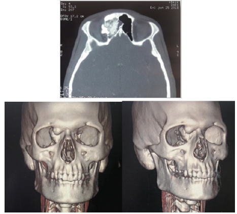
We report the case of an 18 years old male patient, without a prior medical condition, who presents with a mass of the right superior inner angle of the orbit, which grew slowly over a period of one year. There were no other symptoms, particularly no compressive signs such as headaches or diplopia. Clinical examination found a hard mass of the right superior inner corner of the orbit, seeming to be of an osseous nature, measuring approximatively 20 mm in diameter, associated with a slight proptosis of the right eye. The examination of the nasal cavities found no particular anomaly. CT scan imaging revealed a right fronto-ethmoidal osteoma, protruding into the orbit, measuring 45x35x25 mm, repressing the right (Figure 1). The patient benefited from combined endoscopic and open surgery. We performed endoscopic exploration first. The osteoma was discovered in the middle meatus. We then proceeded to the complete resection of the tumor via a supra-brow incision combined to transnasal endoscopic drill cavitation. There were no post-surgery complications, with the disappearance of the tumor and the regression of the proptosis.
Discussion
Osteomas are rare slow-growing, benign bone tumors. It is the most frequent benign tumor of the paranasal sinuses [4,5], affecting in descending order of frequency: the frontal sinuses (50%), the ethmoidal cells (40%), the maxillary sinuses (6%), and the sphenoidal sinuses (4%) [5-7] . Orbital involvement is a rare occurrence, following the extension from the frontal sinus or the ethmoidal cells, that can lead to ocular symptoms [4,8,9]. Our patient had a fronto-ethmoidal osteoma with orbital extension. Osteomas can occur at any age [5], with most cases diagnosed during the 4th and 5th decade of life [10-13] , and there seems to be a slight gender predilection, as 60% of the reported cases are males [10,12,14].
These tumors are often small and asymptomatic, though patients can present with proptosis, diplopia, headaches, nasolacrimal duct obstruction, or even loss of vision [15-17]. More serious complications include intracranial pneumatocele, meningitis, neumoencephalus, subdural abcesses and compressive neuropathies [1,2] . In our case, the patient presented with a process of the right inner angle associated with proptosis, with no other symptoms. Differential diagnosis is made mainly with other bone tumors, such as ossifying fibroma, osteoblastoma, fibrous dysplasia, osteosarcoma, and orbital metastasis [14,18] .
CT scan is considered the golden standard for diagnosis [3,15] . Images show a dense, homogenous mass, with regular limits, arising from paranasal sinuses, well defined, and resembling the cortical bones in its ivory form or with a ground-glass appearance in its spongy form [5]. It is important to note that osteomas are discovered incidentally in 3% of CT scans [13,19-22]. For a small and asymptomatic osteoma, a conservative approach is usually adopted, with regular check-ups and eventually CT scans to survey its growth and extension. As for symptomatic osteomas, two surgical approaches are possible: endoscopic surgery and open surgery. Endoscopic surgery is actually considered to be the treatment of choice for paranasal sinuses osteomas [23-26] . It is a safe and effective technique, offering comestic advantages and lower morbidity rates than open approaches [23,27] .
Endoscopic surgery through an intranasal drill is often sufficient to remove small ethmoidal and frontal osteomas. In case of orbital extension, open surgery might be needed to complete the removal of the tumor after endoscopy is used to drill its center, creating a cavitation of the osteoma [23]. This was the case for our patient, who benefited from intranasal endoscopic drill cavitation first, completed by a suprabrow incision to completely remove the osteoma. There are many complications that may occur after surgery, including diplopia, iatrogenic paralisys, reccurent frontal sinusitis, meningitis, and enophtalmos [2,28] . Additionally, incomplete resection of the osteoma might result in a recurrence. No such complications were observed in our patient after surgery.
Conclusion
Although osteomas are the most frequent tumors of the paranasal sinuses and are often asymptomatic, they can present with a variety of symptoms, and orbital extension is a rare occurrence. CT scan imaging is necessary for the surgeon to make the diagnosis and to define the limits and extension of the tumor. Surgical treatment, when indicated, can resort to two techniques: endoscopic surgery, which is the treatment of reference, and open surgery, though sometimes a combination of both can be necessary
References
- Jack LS, Smith TL, Ng JD (2009) Frontal sinus osteoma presenting with orbital emphysema. Ophthalmic Plast Reconstr Surg 25(2): 155-157.
- Blanco Domínguez I, Oteiza Álvarez AV, Martínez González LM, Moreno García Rubio B, Franco Igle sias G, et al. (2016) Frontoethmoidal osteoma with orbital extension. A case report. Arch Soc Esp Oftalmol 91(7): 349- 352.
- RP Exley, A Markey, S Rutherford, RK Bhalla (2015) Rare giant frontal sinus osteoma mimicking fibrous dysplasia. The Journal of Laryngology & Otology 129(3): 283-287.
- Kim AW, Foster JA, Papay FA, Wright KW (2000) Orbital extension of a frontal sinus osteoma in a thirteen-year-old girl. J AAPOS 4(2): 122-124.
- S Haddar, H Nèji, C Dabbèche, Y Guermazi, K Fakhfakh, et al. (2013) Fronto-orbital osteoma. Answer to the e-quid Unilateral exophthalmos in a 30-year-old man. Diagnostic and Interventional Imaging 94(1): 119- 122.
- M Halhal, M Naciri, O Berbich, W Benabdellah, A Berraho (2000) Osteoma of the orbit. A case report. J Fr Ophtalmol 23(9): 888-891.
- Ersner MS, Saltzman M (1938) Osteoma of the sinusis. Laryngoscope 48: 29-37.
- Becelli R, Santamaria S, Saltarel A, Carboni A, Iannetti G (2002) Endoorbital osteoma: two case reports. J Craniofac Surg 13(4): 493-496.
- Roberto Saetti, Marina Silvestrini, Surendra Narne (2005) Ethmoid osteoma with frontal and orbital extension: Endoscopic removal and reconstruction. Acta Oto-Laryngologica 125(10): 1122-1125.
- James M Mc Cann, Donald Tyler Jr, Robert D Foss (2015) Sino-Orbital Osteoma with Osteoblastoma-Like Features. Head Neck Pathol 9(4): 503-506.
- Nielsen GP, Rosenberg AE (2007) Update on bone forming tumors of the head and neck. Head Neck Pathol 1(1): 87-93.
- Earwaker J (1993) Paranasal sinus osteomas: a review of 46 cases. Skeletal Radiol 22(6): 417-423.
- Tatsuyuki Ishii, Yoshiaki Sakamoto, Tomoru Miwa, Kazunari Yoshida, Kazuo Kishi (2018) A Giant Osteoma of the Ethmoid Sinus. J Craniofac Surg 29(3): 661-662.
- McHugh JB, Mukherji SK, Lucas DR (2009) Sino-orbital osteoma: a clinicopathologic study of 45 surgically treated cases with emphasis on tumors with osteoblastoma-like features. Arch Pathol Lab Med 133(10): 1587-1593.
- Nurdogan Ata, Mesut Sabri Tezer, Ersen Koç, Gültekin Övet, Ömer Erdur (2017) Large Frontoorbital Osteoma Causing Ptosis, The Journal of Craniofacial Surgery 28(1): p.e17-p.e18.
- Ioannis Yiotakis, Anna Eleftheriadou, Evagelos Giotakis, Leonidas Manolopoulos, Eliza Ferekidou, et al. (2008) Resection of giant ethmoid osteoma with orbital and skull base extension followed by duraplasty. World J Surg Oncol 6: 110-210.
- Koktekir BE, Ozturk K, Gedik S, Guzel H, Karabagli P (2012) Giant ethmoido-orbital osteoma presenting with dacryocystitis and metamorphopsia. J Craniofac Surg 23(5): e390–e392.
- Selva D, White VA, O’Connell JX, Rootman J (2004) Primary bone tumors of the orbit. Surv Ophthalmol 49(3): 328-342.
- Leslie A Wei, Nicholas A Ramey, Vikram D Durairaj, Vijay R Ramakrishnan, Augusto V Cruz, et al. (2014) Orbital Osteoma: Clinical Features and Management Options. Ophthal Plast Reconstr Surg 30(2): 168-174.
- Erdogan N, Demir U, Songu M, Ozenler NK, Uluç E, et al. (2009) A prospective study of paranasal sinus osteomas in 1,889 cases: changing patterns of localization. Laryngoscope 119(12): 2355-2359.
- Mansour AM, Salti H, Uwaydat S, Dakroub R, Bashshour Z (1999) Ethmoid sinus osteoma presenting as epiphora and orbital cellulitis: case report and literature review. Surv Ophthalmol 43(5): 413-426.
- Vowles RH, Bleach NR (1999) Frontoethmoid osteoma. Ann Otol Rhinol Laryngol 108(5): 522-524.
- Pons Y, Blancal JP, Vérillaud B, Sauvaget E, Ukkola Pons E, et al. (2013) Ethmoid sinus osteoma: Diagnosis and management. Head Neck 35(2): 201-204.
- Koivunen P, Loppo nen H, Fors AP, Jokinen K (1997) The growth rate of osteomas of the paranasal sinuses. Clin Otolaryngol Allied Sci 22(2): 111-114.
- Trinidade A, Shakeel M, Moyes C, Ram B (2010) The evidence-based management of bilateral ethmoid osteomas: diagnosis, endoscopic resection and review of the literature. West Indian Med J 59(2): 188-191.
- Schick B, Steigerwald C, el Rahman el Tahan A, Draf W (2001) The role of endonasal surgery in the management of frontoethmoidal osteomas. Rhinology 39(2): 66-70
- Gerbrandy SJ, Saeed P, Fokkens WJ (2007) Endoscopic and trans-fornix removal of a giant orbital-ethmoidal osteoma. Orbit 26(4): 299-301.
- Turri Zanoni M, Dallan I, Terranova P, Battaglia P, Karligkiotis A, et al. (2012) Frontoethmoidal and Intraorbital Osteomas Exploring the Limits of the Endoscopic Approach. Arch Otolaryngol Head Neck Surg 138(5): 498-504.


 We use cookies to ensure you get the best experience on our website.