Research Article 
 Creative Commons, CC-BY
Classification of Salivary Gland Diseases among Yemenis: (A Prospective Hospital-Based study)
*Corresponding author: Ali Ali AL-Zamzami, Consultant of oral and maxillofacial surgery, Head of the consultant unit of head and neck surgery, AL-Gemhori Teaching Hospital, Republic of Yemen, Yemen.
Received: July 19, 2019; Published: August 01, 2019
DOI: 10.34297/AJBSR.2019.04.000800
Abstract
Objective: This study aims to classification of salivary gland diseases among Yemenis to established data base, determine the common disease, common site and the relation of these disease to the age and gender.
Material and Methods: The present study designed as a prospective descriptive hospital –based study carried out at Al-gomhori –Teaching Hospital in Sana’a. All patients attending to the department of oral and maxillofacial surgery and to the consultant unit of the head and neck surgery complain of salivary gland disease were examination. Data were collected from patient history (using a questionnaire form), clinical examination of patients, radiographs examination and from the histopathology results of the biopsies. Biopsy result used to confirm the diagnosis.
Results: A total of 140 cases of salivary gland diseases were studied,64 cases (45.7%) were males, and 76 cases (54.3%) were females, male to female ratio was 0.8-1. The age of patients was ranged from 3 to 82 years, with mean age for both gender of 40. 09±21.149. The majority of patients (79%) were over the age of 20 years. The most common disease of salivary glands tumor was salivary gland tumor, accounting 43.6%, followed by cystic lesions 20.7% and sialolithiases (20.0%). The less common salivary gland diseases were sialadenitis and sialadenosis, accounting,12.9% and 2.9% respectively. Sqaumous cell carcinoma was the most common types of salivary gland malignancy, accounting 50. 0%. Pleomorphic adenoma was the most common types of benign tumors accounting,78.3%. Major salivary glands were the most involving site by both tumors. Rhanula was the most common type of salivary gland cyst accounting, 75.9%, usually found on the sublingual glands. Sialolithiases, usually found on the major salivary glands, 89.3% of this disease were located on the submandibular gland. Sialadenitis, frequently caused by bacterial and viral infections, 77% of them were bacterial sialadenitis and 22.2% were viral sialadenitis. Bacterial sialadenitis, commonly located on the submandibular gland, whereas, viral sialadenitis restricted to the parotid glands.
Conclusion: One hundred and forty cases of salivary gland disease among Yemenis were studied,64 cases (45.7%) were males, and 76 cases (54.3%) were females, male to female ratio was 0.8-1. The age of patients was ranged from 3 to 82 years, with mean age for both gender of 40. 09±21.149. The majority of patients (79%) were over the age of 20 years. Salivary glands tumor was the most common salivary gland disease, accounting 43.6%, followed by the salivary gland cyst and sialolithiases accounting, 20.7% and 20.0% respectively. Sqaumous cell was the most common type of salivary gland malignancies accounting 50. 0%. Pleomorphic adenoma was the most common type of benign tumors accounting 78. 3%. Both salivary gland tumors commonly located on the major salivary glands. Rhanula was the most common type of salivary gland cyst, accounting 75.9%,usually located on the sublingual gland. Sialolithiases, accounting 20.0% of all salivary gland disease, 89.3% of this disease were located on the submandibular gland. The less common types of salivary gland disease were sialadenitis and Sialosis, accounting 12.09% and 2.9% respectively.
Keywords: Salivary gland disease; Sialadenitis; Sialolithiases; Salivary gland tumors; Salivary gland cyst
Introduction
Salivary glands divided into major and minor salivary glands. Major salivary glands include three paired of the glands ; parotid, submandibular and sublingual glands. Of minor salivary glands, there are a hundred of these glands’ distribution throughout the mucosal membrane of the upper digestive tract. Saliva is the product of the major and minor salivary glands. It is a highly complex mixture of the water, organic and non-organic components. They also produce enzyme lubrication, mixing agents and immune factors that play an important role of lubrication of the mouth, mastication and swallowing of the food and protection of the oral cavity and teeth [1-4]. Salivary gland disease was related to many causes. The most common causes were; sialadenitis (bacterial and viral infections), sialolithiases (stone formation within the glands or on the duct), sialadenosis (systemic disease), cystic lesions and salivary gland neoplasms. Clinically, salivary gland disease presented as painless or painful swelling on the affected gland. Parotid gland disease presented as swelling below and front of the ear. Submandibular disease presents as swelling on the upper part of the neck. Whereas, sublingual gland and minor salivary gland diseases were presented as submucosal swelling on the oral cavity [5].
Sialadenitis or salivary glands infections, frequently caused by bacterial or viral infections. Bacterial sialadenitis most commonly caused by staphylococcus micro-organisms that usually found normally within the oral cavity [2]. It’s most commonly related to the chronic reduction of the salivary flow that lead to diminished mechanical flushing, which allow bacteria to colonize the oral cavity and then invaded the salivary glands (retro-grad infection) [6-8]. Bacterial sialadenitis divided into acute and chronic sialadenitis. Acute sialadenitis presented as a redness and tenderness swelling of the affected gland, whereas, chronic sialadenitis presented as painless swelling and in sometimes associated with pus discharged [5]. Mumps was the most common type of viral sialadenitis, commonly caused by RNA paramyxovirus, which transmitted by direct contact with salivary droplets. Mumps usually presented as acute painful swelling of the parotid gland, frequently bilateral. Children are most affected with peak incidence occurring at approximately 4 to 6 years of the age [2,9]. Sialolithiases or stone formation within the salivary glands or on the ducts occur frequently on submandibular salivary gland. Exact causes of stone formation are unknown, it’s felt to be ; secondary to the stagnation of saliva, partial obstruction of the salivary gland duct, trauma, dehydration, medication effects (such as diuretic and anticholinergic agents) and smoking [10]. Sialolithiasis presented as painless swelling or may be noted incidentally during physical or radiograph examinations. Occasionally, it’s can cause painful swelling or progress to acute sialadenitis [10,11]. Sialolithiases more commonly affects adults in their 3rd to 6th decades of life, it can find also in children. Submandibular gland and it’s duct were the most affected site, accounting 80%, followed by the parotid gland 20%. Sublingual gland and minor salivary glands are rarely affected [12-14].
Sialadenosis or Sialosis defined as a bilateral, persistent, nonneoplastic, non-inflammatory painless swelling of the salivary glands, more commonly on the parotid glands [2,15]. Sialadenosis occurs most commonly in relation to alcoholism but can develop in relation to many systemic disorders such as ; diabetes mellitus, malnutrition and even idiopathic disease [16-18]. Rhanula and mucocele are the most common type of salivary gland cysts. Both types classified as extravasation and retention cyst. Rhanula, defined as a cystic lesion of the salivary glands, most commonly on the sublingual gland, occur due to extravasation or retention of mucous from sublingual gland or duct due to trauma or obstruction. It’s classified into superficial and deep rhanula. Superficial rhanula, appear as a bluish lesion on the anterior part of the floor of the mouth. Whereas, deep rhanula occurs when the sublingual salivary gland lobe and its duct penetrates through the floor of the mouth muscles and present as extraoral mass (plunging rhanula) on the submandibular or submental regions [2,19,20]. Mucocele is a clinical term that describes minor salivary gland cyst, that appear as a bluish swelling, more commonly on the lower lip, buccal mucosa and ventral surface of the tongue. It’s caused by the accumulation of the saliva at the site of a traumatized or obstruction minor salivary gland duct [19]. Salivary gland tumors are specific neoplasms in the oral and maxillofacial region and accounts for about 3 - 6 % of all head and neck tumors. It’s more frequent in adult than in children, the maximum age of incidence is the 4th decade of life for benign tumors and 5th decade for malignant tumors [21-24].
Pleomorphic adenoma was the most frequently type of benign tumor, accounts for about 64. 0 %, followed by Wharton’s tumor and haemangioma, accounting 4. 8% and 2. 4% respectively. Of malignant tumors, adenoid cystic carcinoma was the most common type of salivary gland malignancies, followed by mucoepidermoid carcinoma and carcinoma in expleomorphic adenoma [25]. Other study showed that, Squamous cell carcinoma, was the most common type of salivary gland malignancies, followed by mucoepidermoid carcinoma and adenoid cystic carcinoma [26]. Both types of salivary gland tumors, present as a painless mass in the affected gland. Findings that are concerning for malignancy include ; pain, facial paresis, fixation of the mass to the skin or underling tissues and palpable neck- lympho-adenopathy [27-30]. Most salivary gland tumors (80%) occurring in the parotid gland, the remaining 20% occur in the submandibular gland, sublingual gland and minor salivary glands. The present study aimed to studied of salivary gland diseases among Yemenis to determine the common disease, common site, the relation of these diseases to the age and gender of the patients and to comparison the provides data with previously published studies from different geographical sites.
Material and Methods
This study design as a prospective, descriptive hospital based –study, carried out at AL-gomhori Teaching Hospital in Sana’s Republic of Yemen. The material consisted 140 of Yemeni patients who attending to the Department of Oral and Maxillofacial Surgery and to the Consultant Unit of the Head and Neck Surgery and who were diagnosed clinically and radiographically as having salivary gland diseases. Histopathological examination used to confirm the diagnosis. Data were collected from, patient history (using a questionnaire sheet), clinical examination of the patients, radiograph examinations (plain radiographs, sialographs, ultrasonograph, and CT-Scan) and histopathological examination (Fin Needle Aspiration biopsy and excisional biopsy). Histopathological results were used to confirm the diagnosis. Data were analysis using SPSS programversion 24. Results were presented as simple frequencies and descriptive statistics. Pearson’s Chi-Square Test was used to assess the association and level of significance among categorical variables, P-value less than 0. 05 is considered as statically significant.
Ethical clearance
The respondent was adequately informed about all relevant aspects of the study including; the aim of the study, the need of investigations, and regular follow up. Privacy of patient is the most of our priority.
Results
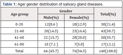
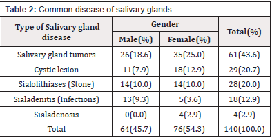
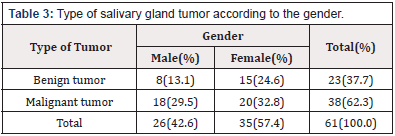
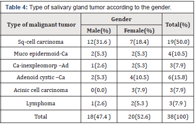
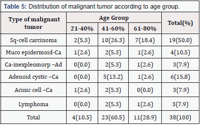
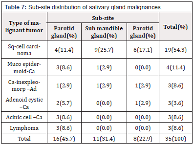
A total of 140 cases of salivary gland diseases were studied,64 cases (45.7%) were males, and 76 cases (54.3%) were females, male to female ratio was 0.8-1. The age of patients was ranged from 3 to 82 years, with mean age for both gender of 40. 09+21.149. The majority of patients (79%) were over the age of 20 years. The peak age of occurrence (35.7%) was between the fourth and sixth decades of life (Table 1). The most common disease of salivary glands was salivary gland tumor, accounting (43.6%), followed by, salivary gland cyst and sialolithiases, accounting 20.7% and 20.0 % respectively. The less common disease were sialadenitis and sialadenosis, accounting 12.9% and 2.9% respectively (Table 2). Of salivary gland tumors,61 cases were reported, 26 cases (42.6%) were males and 35 cases (57.4%) were females, male to female ratio was 0.7-1. Thirty-eight cases (62.3%) of salivary gland tumors were malignant tumors and 32 cases (37.7%) were benign tumors (Table 3) Of malignant tumors, 38 cases were reported, 18 cases (47.4%) were males and 20 cases (52.6%) were females. Male to female ratio was 0.9-1. The majority of patients (89.4%) were over the age of 40 years. The peak occurrence (60.5%) was in between 4th and 6th decades of life (Table 4). Squamous cell carcinoma was the most common type, accounting (50.0%), followed by adenoid cystic carcinoma and muco epidermoid carcinoma, accounting (15.8% and 10.5% respectively). The less common types were, cainexpleomorphic adenoma, acinic cell carcinoma and lymphoma accounting 7.9% for each one (Table 5). More than 92 percent of salivary gland malignancies found on the major salivary glands, 45.7% of these were found on the parotid glands, 31.4% on the submandibular gland and 22. 9% on the sublingual glands (Table 6).
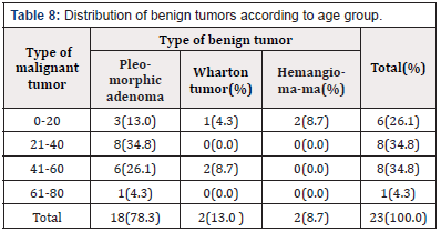
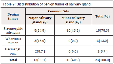
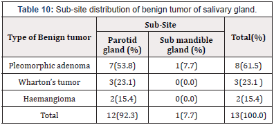
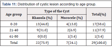
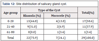
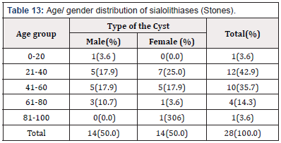
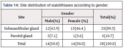
Of benign tumors, 23 cases were reported, 8 cases were males and 15 cases were females, male to female ratio was 0.5:1. The majority of patients (70%) were found between the 3rd and 6 th decades of life. Pleomorphic adenoma was the communist type, accounting (78.3%), followed by Wharton’s tumor and hemangioma accounting (13.0% and 8.7% respectively) (Table 8). Thirteen cases of benign tumors (59.1%) were found on the major salivary glands and 9 cases (40.9%) on the minor salivary glands (Table 9). Ninety two percent of benign tumors of the major salivary gland’s tumors were located on the parotid glands, 53.8 % of these tumors were pleomorphic adenoma, 23. 1% were Wharton’s tumors and 15. 4 % were haemingioma (Table 10). Of cystic lesion,29 cases were reported, 11 cases (37.9%) were males and 18 cases (62.1%) were females, male to female ratio was 0.6:1. The majority of patients (96.5%) were found between the 1st to 4th decades of life, the peak age of occurrence (58.6%) was in the 1st and 2nd decades of life (Table 11). Rhanula and mucocele were reported, accounting 75.9% and 24.1% respectively. Major salivary glands (sublingual glands) were the most affected site (75.9%) (Table 12). Of sialolithiases (or stone formation) 28 cases were seesn, 14 cases were males and 14 cases were females, male to female ratio was I:1. More than (90%) of patients were found over the age of 20 years. Patients on the middle age were commonly affected(78.6%) (Table 13). Submandibular gland was the most common affected site, accounting (89.3%), followed by parotid glands (10. %) (Table 14).
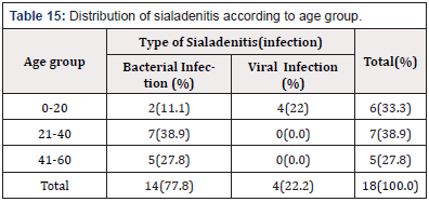
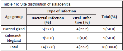
Of sialadenitis (or salivary gland infections),18 cases were reported, 13 cases (72.2%) were males and 5 cases (27.8%) were females, male to female ratio was 2.6:1 (Table 1). Patient age was ranged from 3 to 82 years with mean age of 42. 5±SD. Fourteen cases of sialadenitis (77.8) were bacterial sialadenitis and 4 cases (22.2%) were viral sialadenitis (Table 15). The majority of bacterial sialadenitis (64.3%) were found on the Submandibular gland and the residual percentage 35.7% on the parotid gland. Whereas, viral sialadenitis restricted to the parotid gland (Table 16). Sialadenosis (non-neoplastic, non-inflammatory disease of the salivary glands which commonly related to some systemic disease or disorder) was the less common type, 4 cases were reported accounting 2. 9% of all salivary gland disease. All cases were found in female patients.
Dissection
In the present study, salivary gland tumors were the most common type of salivary gland diseases, 61 cases were reported, 38 cases (62.3%) of them were malignant tumors and 23 cases (37.7%) were benign tumors. Of malignant tumors, males were affected less than females, male to female ratio was 0.9:1. Patients age ranged from 26 to-80 years, the average of the age was 53 years. The majority of patients (89.4%) were over the age of 40 years. These findings are in agreement nearly to many literatures studies that showed a higher frequency of salivary gland malignancy among females, with male to female ratio was 1:1. 8. These studies also showed that, salivary gland malignancy most commonly found over the age of 40 years and the mean age of patients were distributed between, 40 years, 41.38 years, 46 years and 48 years respectively [31,35]. Our study also showed that, Squamous cell carcinoma was the most common type, accounting 54.3%,followed by muco-epidermoid carcinoma 11. 4% and carcinoma- inexpleomorphic adenoma 8.6%. Same findings were reported by Ali AL-Zamzami and Ahmed Suleiman [26] who founds that, sqaumous cell carcinoma was the most common histological type of salivary gland malignancies, followed by mucoepidermoid carcinoma and carcinoma in ex-Pleomorphic adenoma. On the other hand, the prevalence of salivary gland malignancies was various. Many literature studies have reported, adenoid cystic carcinoma [35], carcinoma inexpleomorphic adenoma [36], sqaumous cell carcinoma [37] as the most common type of salivary gland malignancies.
Our findings also showed, the majority of salivary gland malignancies (92%) were located on the major salivary glands, 45. 7% of these tumors were located on the parotid gland, 31.4% on the submandibular gland and 22. 9% on the sublingual gland. Similar observations were reported by Ahmed Suleiman et al. [31] who founds that, major salivary glands (parotid gland and submandibular gland) were the most affected sites with salivary gland malignancies. The above findings were confirmed by Mohammed Isa Kara [25] who found that, parotid gland and submandibular gland were the main affected sites, accounting 61.6% and 16% respectively. Of benign tumors, our study showed, males were affected less than females, male to female ratio was 0.5:1. Patients age was ranged from 3- 82 years with average of 42.5 years. The majority of patients (70%) were over the age of 20 years. Pleomorphic adenoma was the most common type, accounting 77.3% followed by Wharton’s tumors and hemangioma accounted 13. 6% and 9. 1% respectively. Major salivary gland was the most affected site and 90 % of these tumors were located on the parotid gland. Our findings are nearly agreement to literature studies that showed that, males were affected less than females, male to female ratio was 1 : 1. 6. Patient age was ranged from 1 – 88 years with median age of 45 years [30,38]. Other studies showed that, Pleomorphic adenoma was the most common type of benign salivary gland tumors, accounting 64 %, followed by Wharton,’ tumor and hemangioma, accounting 4. 8% and 2. 4% respectively. These studies also showed that, partied gland was the main affected site [21,23,24,32-35].
In our study,rhanula and mucocele were reported. Rhanula was the most common type, accounting 75.9%, the second type was mucocele, accounting 24.1%. Male were affected less than females, male to female ratio was 0.6 –1. Patients age was ranged from 1 to 35 years, the mean age was 18 years±sd. The majority of patients (93%) were found below the age of 35 years. . Major salivary glands (sublingual glands) were the most affected site (75.9%). These findings are similar to many previous studies that showed, salivary gland cyst usually occurred in children and adults with the peak frequency in the second decade of life, the mean age was 18. 5 years. Males were affected less than females, male to female ratio was 1:1.4. These studies also showed that, rhanula was the most common type of salivary gland cyst and frequently found on the major salivary glands, particularly sublingual salivary gland p [37,39-41]. Of sialolithiases or salivary gland stone. This study showed, males and females were equal affected, male to female ratio was 1 : 1. Patients age was running between 19 and 82 years, the mean age was 47. 25 years±SD. The majority of patients (90%) were found over the age of 20 years. All cases of sialolithiases were found on the major salivary glands, 89.3% of these cases were found on the submandibular gland and 10. 7% were found on the parotid gland. Similar findings were reported by Lustman J et al. [13] who founds that, sialolithiases or salivary gland stone can occur in any age, more commonly on the adults. Both sexes were equal affected. Most cases 83% were located on the submandibular gland and 10% on the parotid gland. Other study is coinciding with the above findings. It’s showed that, sialolithiases or salivary gland stone can be found in children, but more commonly on the adults in their third to sixth decades of life. Over 80% of salivary stones occur in the submandibular gland and less than 20 % occurs in the parotid gland [14].
In the current study, sialadenitis (salivary gland infection) occur in males more than females, male to female ratio was 2.6:1. Patient age was ranged from 3 to 82 years; the mean age was 42.5±SD. More than 77% of sialadenitis were bacterial sialadenitis and 22.2% were viral sialadenitis. Bacterial sialadenitis founded on the submandibular gland and parotid gland which represented 64.3% and 35.7% respectively. Whereas, viral Literature studies showed that, sialadenitis occur in males more than females. patient age was ranged from 14-58 years, the majority of patients were found between 4th and 6th decades of life. These observations also showed that, sialadenitis usually caused by bacterial and viral infection, more commonly occur on the major salivary gland(58. 2% on the submandibular gland and 41.8% on the parotid gland [42-44].
Conclusion
Salivary gland disease among Yemenis was studied,140 cases were reported,64 cases (45.7%) were males, and 76 cases (54.3%) were females, male to female ratio was 0.8-1. The age of patients was ranged from 3 to 82 years, with mean age for both gender of 40.09±21.149. The majority of patients (79%) were over the age of 20 years. Salivary gland tumor was the most common disease of salivary glands, accounting (43.6%), followed by salivary gland cyst, sialolithiases, sialadenitis and sialadenosis, accounting (20.7%), (20.0%),(12.9%) and (2.9%) respectively. Sqaumous cell carcinoma was the most common type of salivary gland malignancy, accounting 50.0%. Pleomorphic adenoma was the most common type of benign tumors, accounting 78.3%. Both salivary gland tumors commonly located on the major salivary glands. Rhanula was the most common type of salivary gland cyst, accounting 75.9%. Sublingual gland was the most affected site (75.9%). Sialolithiases (or salivary gland stone) usually located on the major salivary gland and 89.3% of them were found on the submandibular gland. Sialadenitis (or salivary gland infection), frequently caused by bacterial and viral infections, 77% of sialadenitis were bacterial sialadenitis and 22.2% were viral sialadenitis. Bacterial sialadenitis, commonly located on the submandibular gland and less commonly on the parotid gland. Whereas, viral sialadenitis restricted to the parotid glands. Sialadenosis (non-neoplastic, non-inflammatory disease of the salivary glands) was the less common type of salivary gland disease, accounting 2.9% and found only in female patient
References
- TT Senthilnathan, S Thirunavukkarasu (2017) A Clinical Study of Salivary Gland Swelling. Journal of Dental and Medical Sciences (IOSRJDMS) 16(7): 53-57.
- Bova R, St Vincent’s (2006) A guide to salivary gland disorders. Medicine Today 7(2).
- RHB Jones, GJ Findlay (2013) The management of benign salivary disease : a case series. Australian Dental Journal 58: 112-116.
- Baum BJ (1993) Principle of saliva secretion. Ann N Y Acad Sci 694: 17- 23.
- Hisham Mehanna, Andrew McQueen, Max Robinson, Vinidh Paleri (2012) Salivary gland swellings. BMJ 345: e6794.
- Fox PC (1991) Bacterial infections of salivary glands. Curr Opin Dent 1: 411-414.
- Raad II, Sabbagh MF, Caranasos GJ (1990) Acute bacterial sialadenitis : a study of 29 cases and review. Rev Infect Dis 12(4): 591-601.
- Goldberg MH, Bevilacqua RG (1995) Infections of salivary glands. Oral Maxillofac Surg Clin North Am 7(3): 423-430.
- Caplan CE (1999) Mumps in the era of vaccines. Can Med Assoc J 160(6): 856-866.
- Yousem DM, Kraut MA, Chalian AA (2000) Major salivary gland imaging. Radiology 16:19-29.
- Cummings CW (2010) Cummings otolaryngology-head and neck surgery (5th Edn.) Philadelphia, USA pp. 1151-1161.
- Leung AK, Choi MC, Wagner GA (1999) Multiple sialoliths and a sialoliths of unusual size in the submandibular duct : a case report. Oral Surg Oral Med Oral pathol Oral Radiol Endod 87(3): 331-333.
- Lustman J, Regev E, Melamed Y (1990) Sialolithiasis. A survey on 245 patients and a review of the literature. Int J Oral Maxillofac Surg 19(3): 135-138.
- Bodner L (2002) Giant salivary gland calculi : Diagnosis imaging and surgical management. Oral Surg Oral Med Oral Pathol Oral Radiol Endod 94(3): 320-323.
- Rahuram Sivasubramaniam, Siamak Choroomi, Hilton E Stone (2015) Sialolithiasis in a Remnant Wharton’s Duct : A Case Study and Discussion. Journal of Otolaryngology-ENT Research 2(1): 0-12.
- Mandel L, Vakkas J, Saqi A (2005) Alcoholic Sialosis. J Oral Maxillofac Surg 63(6): 402-405.
- Chilla R (1981) Sialadenosis of the salivary glands of the head : studies on the physiology and patho-physiology of parotid secretion. Adv Otorhinolaryngol 26: 1-38.
- Scully C, Bagan JV, Eveson JW (2008) Sialosis : 35 cases of persistent parotid swelling from two countries. Br J Oral Maxillofac Surg 46(6): 468-472.
- Wilcox JW, Hickory R (1978) Nonsurgical resolution of mucoceles. J Oral Surg 36(6): 478.
- Zhao YF, Jia Y, Chen XM, Zhang WF (2004) Clinical review of 580 ranulas. Oral Surg Oral Med Oral Pathol Oral Radiol Endod 98(3): 281-287.
- Li LJ, Li Y, Wen YM, Liu H, Zhao HW (2008) Clinical analysis of salivary gland tumor cases in West China in past 50 years. Oral Oncolo 44: 187- 192.
- Minicucci EM, De Campos EB, Weber SA, Domingues MA, Ribeiro DA (2008) Basal cell carcinoma of the upper lip from minor salivary gland origin. Eur J Dent 2: 213-216.
- Wang D, Li Y, He H, Liu L, Wu L, et al. (2007) Intraoral minor salivary gland tumors in a Chinese population : a retrospective study on 737 cases. Oral Surg Oral Med Oral Pathol Oral Radiol Endod 104(1): 94-100.
- Dhanuthai K, Sappayatosok K, Kongin K (2009) Pleomorphic adenoma of the palate in a child : a case report. Med Oral Patol Cir Bucal 14(2): E73-75.
- Muhammed Isa Kara, Fahrettin Goze, Seref Ezirganh, Serkan Polat, Suphi Muderris et al. (2010) Neoplasms of the salivary glands in a Turkish adult population. Med Oral Patol Oral Cir Bucal 15(6): e880-5.
- Al-Zamzami A, Suleiman AM (2018) Oral cancer among Yemenis : A prospective hospital based-study. Dent Med Res Journal 6: 32-36.
- To VS, Chan JY, Tsang RK, Wei WI (2012) Review of salivary gland neoplasms. ISRN otolaryngology.
- Akar HH, Patiroglu T, Duman L (2014) A selective IgA deficiency in a boy who presented recurrent parotitis. Eur J Microbiol Immunol(Bp) 4: 144- 146.
- Main JH, Orr JA, McGurk FM, McComb RJ, et al. (1975) Salivary gland tumors review of 643 cases. J Oral Pathol Med 5: 88-102.
- Torabinia N, Khalesi S (2014) Clinicopathological study of 229 cases of salivary gland tumors in Isfahan population. Dent Res J(Isfahan) 11: 559-563.
- Ahmed S, Yousif OY, Abuzeid M (2018) Tumors of Salivary Glands in Sudan. Inter J Otorhino-laryngology 5(1): 5.
- Suphashraj K (2008) Salivary gland tumors a single institution experience in India. Br J Oral Maxillofac Surg 46(8): 635-638.
- Al-Khateeb TH, Ababneh KT (2007) Salivary gland tumors in north Jordanians a descriptive study. Oral Surg Oral Med Oral Pathol Oral Radiol Endod 103:e53-9.
- Vargas PA, Gerhad R, Araujo Filho VJ, De Castro (2002) Salivary tumors in Brazilian population: a retrospective study of 124 cases. Rev Hosp Clin Fac Med Sao Paulo 57: 271-276.
- Kamulegeya A, Kasangaki A (2004) Neoplasms of the salivary glands: a descriptive retrospective study of 142 cases-Mulago Hospital Uganda. J Contemp Dent Pract 5(3):16-27.
- Eveson JW, Cawson RA (1985) Salivary gland tumors.A review of 2410 cases with particular reference to histological types, site, age and sex distribution. J Pathol 146: 51-58.
- Otoh EC, Johnson NW, Olasoji H, Danfillo IS, Adeleke OA (2005) Salivary gland neoplasms in Maiduguri, north-eastern Nigeria. Oral Dis 11: 386- 391.
- Jaber MA (2006) Intraoral minor salivary gland tumors: a review of 75 cases in a Libyan population. J Oral Maxillofac Surg Med Pathol 35(2): 150-154.
- Moosa Bahnassy (2005) Ahuge Oral Ranula. Oman Medical Journal 24(4).
- Daniel Kokong, Augustin Idah, SuleAugustin (2017) Ranula: Current Concept of Pathophysiologic Basis and Surgical Management Option. World J Surg 41(6): 1476-1481.
- Sigismunt PE, Bozzato A, Schuman M (2013) Management of ranula : 9 years, clinical experience in pediatric and adult patients. J Oral Maxillofac Surg.2013;7(3): 538-544.
- Christopher G, Pace, Kyung-Gyun Hwang, Maria Papadaki, Maria J (2014) Interventional Sialoendoscopy for Treatment of Obstructive Sialadenitis. J Oral Maxillofac Surg 72: 2157-2166.
- Krause CH, Eastick K (2006) Real time PCR for mumps diagnosis on clinical specimens. J Clin Viro 37(3): 184-189.
- Rajiv Kumar, Amit Kumar, Renu Sawal (2012) Sialadenitis-A Salivary gland disease. International Journal of Pharmaceutical Erudition 1(4): 16-240.




 We use cookies to ensure you get the best experience on our website.